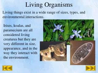
Living organisms verses inanimate objects
Living organisms verses inanimate objects. Characteristics of living organisms. Maintaining life processes. Normal vital functions Body systems operating to obtain oxygen, nutrients, Responding to environment and adapt to environmental stimuli Maintaining life in disease Treatments

Living organisms verses inanimate objects
E N D
Presentation Transcript
Living organisms verses inanimate objects Characteristics of living organisms
Maintaining life processes • Normal vital functions • Body systems operating to obtain oxygen, nutrients, • Responding to environment and adapt to environmental stimuli • Maintaining life in disease • Treatments • Biomedical Devices
First do no harm Supporting life processes
What are characteristics of life? • ___________________ • ___________________ • ___________________ • ___________________ • ___________________
Microbes can cause disease in animals The microorganisms shown here are: A, the bacterium Escherichia coli; B, a photosynthetic cyanobacterium; C, a fungus; D, Ebola virus; E, the protozoan malaria parasite. (Sources: B, Mike Clayton; C-E, CDC). Note that the scale on each of these pictures is different.
Infectious disease • In the U.S., infectious diseases have been significantly diminished over the last 150 years (small pox, dysentery, leprosy, etc.) • Developing nations are still battling disease problems (Haiti) • New diseases have appeared (AIDS) that present different problems
Microorganisms • Microbes make up the major portion of the biomass present on the Earth. Therefore, the nutrients they eat and the products they form greatly influence the environment. • Cyanobacteria and algae in the oceans are responsible for most photosynthesis and are a major sink for carbon dioxide, a greenhouse gas. • Microbes release nutrients from dead organisms, making them available to the rest of the ecosystem. • Some microbes play a role in the production of energy, while other microbes interfere with energy production.
Helpful and harmful Microorganisms played an important role in removing many of the pollutants released during the Exxon Valdez oil spill in Prince William Sound. Interestingly, microbes were not added to the site, but the clean-up relied on bacteria from that environment. A nutrient solution was sprayed onto the oil to encourage the growth of oil-degrading microbes. Though this was one of the more successful methods used to clean up the oil, but no treatment removed all of the pollutants.
What is disease? • Ignaz Semmelweis showed that child-bed fever was spread by physicians and could be prevented by careful hand washing with chloride of lime (1861). • Louis Pasteur, while working on sour wine, discovered that unwanted microbes were infecting the wine. He correctly deduced that infectious disease was caused by similar infections with harmful microbes (1865). • Robert Koch was the first to isolate a disease-causing microbe, Bacillus antrhacis. In the process he developed techniques and standard protocols for defining the cause of a disease (1876).
Robert Koch (1876) • A set of rules for the assignment of a microbe as the cause of a disease: • The specific organism should be shown to be present in all cases of animals suffering from a specific disease, but should not be found in healthy animals. • The specific microorganism should be isolated from the diseased animal and grown in pure culture on artificial laboratory media. • This freshly isolated microorganism, when inoculated into a healthy non-immune laboratory animal, should cause the same disease seen in the original animal. • The microorganism should be reisolated in pure culture from the experimental infection.
FUNDAMENTALS • Microbes are useful tools in research because of their rapid life cycle, their simple growth requirements, and their small size. • Due to this simplicity, microbes have been essential in understanding core questions in biology. • Attempts to classify microorganisms have lead to a classification system that divides all organisms into three domains of life: Archaea, Bacteria, and Eukarya. • Microbes provide tools for use in molecular biology. These tools have allowed scientists to make rapid progress in investigating many types of microorganisms.
Prokaryote and Eukaryote Cells • Both have DNA as their genetic material (it’s DNA that tells cells what kind of cells they should be). • Both are covered by a cell membrane. • Both contain RNA. • Both are made from the same basic chemicals: carbohydrates, proteins, nucleic acid, minerals, fats and vitamins. • Both have ribosomes (the structures on which proteins are made). • Both regulate the flow of the nutrients and wastes that enter and leave them. • Both have similar basic metabolism (life processes) like photosynthesis and reproduction. • Both require a supply of energy. • Both are highly regulated by elaborate sensing systems ("chemical noses”) that make them aware of the reactions within them and the environment around them.
Differences • Eukaryotic cells contain two important things that prokaryotic cells do not: a nucleus and organelles (little organs) with membranes around them. • DNA arrangementAlthough both eukaryotic and prokaryotic cells contain DNA, the DNA in eukaryotic cells is held within the nucleus. In prokaryotic cells, the DNA floats freely around in a unorganized manner. • Presence of organellesThe organelles in eukaryotic cells allow them to perform more complex functions than prokaryotic cells, which donot have these little organs. • Some of the organelles in eukaryotic cells are: • The Nucleus – the “brain” or control center of the cell. It contains DNA, which makes up genes. That DNA gets transcribed, or copied onto messenger RNA. That messenger carries a copy of the genes orders for certain protein production. These orders go to the protein factories. • Ribosomes – These are the protein factories. They follow instructions from messenger RNA (remember that the messenger RNA got its orders from the DNA). The instructions tell the ribosomes to make specific proteins. Note, this particular organelle is found in prokaryotes too! • Endoplasmic Reticulum (ER) – structures that modify proteins produced in the ribosomes. Not all of the proteins made by the ribosomes need changing, but those that do get “altered” here. • Golgi Apparatus – This structure will make even more changes to the proteins that already got changed when they were in the E.R. Remember those proteins were made in the ribosomes, changed once in the E.R. and will be changed again in the Golgi Apparatus. The Golgi also acts as a post office by packaging and shipping proteins to other parts of the cell or out of the cell. • Mitochondria – structures which produce the cell’s energy, a.k.a. powerhouses of the cell. • Chloroplasts – structures which allow plants to trap sunlight and carry out photosynthesis.
Eukaryotes • Size Eukaryotic cells are, on average, ten times larger than prokaryotic cells. • Cell Wall DifferencesProkaryotic cells have a cell wall composed of peptidoglycan (amino acid and sugar). Some eukaryotic cells also have cells walls, but none that are made of peptidoglycan. • Flagella ArrangementThe flagella in eukaryotic cells are different from the flagella in prokaryotic cells. Flagella are the structures that help cells move (scientists call it motility). The flagella in eukaryotic cells are composed of several filaments and are far more complex than the flagella in prokaryotic cells. • All cells have their genes arranged in linear chains called chromosomes, but eukaryotic cells contain two (or more) copies of every gene. During reproduction, the chromosomes of eukaryotic cells undergo an organized process of duplication called mitosis.
Prokaryotes or Procaryotes • Groups of organisms that lack cell nucleus • They do not have true nuclei containing DNA • Prokaryotes have a larger surface area to volume ratio giving them a higher metabolic rate, a higher growth rate and consequently a shorter generation time when compared to prokaryotes. Typical cell structure of a eubacterium
The Procaryotes Eubacteria Archaebacteria
Prokaryotes • Typically envisaged to be unicellular but are capable of forming stable aggregate communities • Communities are often encased in a stabilizing polymer matrix (slime) called biofilms, capable of doing cell to cell signaling • Originally in biology, the distinction between prokaryotes and eukaryotes were so great that it was the basis for a 2 tier grouping for cells • A criticism of this classification is that the word "prokaryote" is based on what these organisms are not (they are not eukaryotic), rather than what they are (either archaea or bacteria) • This arrangement of Eukaryota (also called "Eukarya"), Bacteria, and Archaea is called the three-domain system replacing the traditional two-empire system
Gram positive and gram negative bacteria • Gram positive and gram negative refers to how a bacteria reacts to a gram stain. If it takes the initial stain, it will be purple and be considered gram positive. If it doesn't take the initial stain, it will be pink and gram negative. The difference is the outer casing of the bacteria. A gram positive bacteria will have a thick layer of peptidoglycan (a sugar-protein shell) that the stain can penetrate. A gram negative bacteria has an outer membrane covering a thin layer of peptidoglycan on the outside. The outer membrane prevents the initial stain from penetrating.
Gram Positive or negative • There are two distinct types of bacteria based on the structural differences of their cell walls. Gram-positive bacteria will retain the crystal violet dye when washed in a decolorizing solution. The pathogenic capability of Gram-negative bacteria is often associated with certain components of Gram-negative cell walls, in particular the lipopolysaccharide (also known as LPS or endotoxin) layer.[1] In humans, LPS triggers an innate immune response characterized by cytokine production and immune system activation. Inflammation is a common result of cytokine (from the Greek cyto, cell and kinesis, movement) production, which can also produce host toxicity.
Gram negative • The proteobacteria are a major group of Gram-negative bacteria, including Escherichia coli, Salmonella, Shigella,and other Enterobacteriaceae, Pseudomonas, Moraxella, Helicobacter, Stenotrophomonas, Bdellovibrio, acetic acid bacteria, Legionella and alpha-proteobacteria as Wolbachia and many others. Other notable groups of Gram-negative bacteria include the cyanobacteria, spirochaetes, green sulfur and green non-sulfur bacteria. • Medically relevant Gram-negative cocci include three organisms, which cause a sexually transmitted disease (Neisseria gonorrhoeae), a meningitis (Neisseria meningitidis), and respiratory symptoms (Moraxella catarrhalis). • Medically relevant Gram-negative bacilli include a multitude of species. Some of them primarily cause respiratory problems (Hemophilus influenzae, Klebsiella pneumoniae, Legionella pneumophila, Pseudomonas aeruginosa), primarily urinary problems (Escherichia coli, Proteus mirabilis, Enterobacter cloacae, Serratia marcescens), and primarily gastrointestinal problems (Helicobacter pylori, Salmonella enteritidis, Salmonella typhi). • Gram-negative bacteria associated with nosocomial infections include Acinetobacter baumannii, which cause bacteremia, secondary meningitis, and ventilator-associated pneumonia in intensive care units of hospital establishments. Gram-negative Pseudomonas aeruginosa bacteria (pink-red rods).
Treatment • The following treatments are recommended: • Azithromycin 1 gram oral as a single dose, or • Doxycycline 100 milligrams twice daily for seven to fourteen days. • Tetracycline • Erythromycin
Gram positive • Gram-positivebacteria are those that are stained dark blue or violet by Gram staining. This is in contrast to Gram-negative bacteria, which cannot retain the crystal violet stain, instead taking up the counterstain (safranin or fuchsin) and appearing red or pink. Gram-positive organisms are able to retain the crystal violet stain because of the high amount of peptidoglycan in the cell wall. Gram-positive cell walls typically lack the outer membrane found in Gram-negative bacteria. Gram stain for anthrax
Gram positive bacteria • The following characteristics are generally present in a Gram-positive bacterium:[2] • cytoplasmic lipid membrane • thick peptidoglycan layer • teichoic acids and lipoids are present, forming lipoteichoic acids which serve to act as chelating agents, and also for certain types of adherence. • capsule polysaccharides (only in some species) • flagellum (only in some species)
Treatment • Studies have also shown that the broader-spectrum of antibiotics offer more effective short treatment courses than the traditional 10 days of Penicillin V,[30] but noted that "widespread use of broad-spectrum agents for a common infection is a significant concern in an age of increasing bacterial antibiotic resistance".[31] It is important to complete the full course of antibiotics to prevent rheumatic fever or an abscess on the tonsils. In one report of 500 patients, 30% had group A beta-hemolytic streptococcal pharyngitis, 0.2% had rheumatic fever and 0.2% had peritonsillar abscess (an abscess on the tonsils).[6] • Azithromycin and other macrolides have been used to treat strep throat in penicillin-sensitive patients, however macrolide resistant strains of GAS are not uncommon. In these strains, cross-resistance to macrolides, lincosamides, and streptogramins is possible.
Experiments for Home and Classroom • This site provides an excellent description of prokaryotic and eukaryotic cells and allows students to conduct "cell learning" experiments using common household objects such as Tupperware™ containers, Ziploc™ bags, applesauce, marshmallows, spaghetti and breakfast cereal. Click: http://stars.eng.usf.edu/genetic%20engineering%20module/TheCellPowerHouse.doc • This site offers virtual tours of the component parts (the structure and organelles) of both kinds of cells. It also contains a great deal of interesting material presented in an easily understandable way. Go to this site and click on the topic that interests you or your students most:http://www.cellsalive.com/cells/3dcell.htmIn this activity, students are invited to create a booklet explaining cell theory, the function of organelles and cell membrane processes. Materials needed include index cards, pictures of cellular organelles, yarn or string and markers.There are both online and printable versions. Click: http://micro.magnet.fsu.edu/cells/plantcell.html
The protists or protozoans Malaria is caused by four species of protozoan parasites Plasmodium falciparum Plasmodium vivax Plasmodium ovale Plasmodium malaria Trichomonads
Budding yeast • Beer! • Bread • Candidiasis – sometimes called thrush or yeast infections, can be very serious in immunodeficient patients
Plants – cells contain chloroplasts and carry out photosynthesis, i.e. convert sunlight CO2 and H20 to sugars
Animals Invertebrates – do not have a backbone Vertebrates do have a backbone
ATP • Adenosine-5'-triphosphate (ATP) is a multifunctional nucleotide used in cells as a coenzyme. It is often called the "molecular unit of currency" of intracellular energy transfer.[1] ATP transports chemical energy within cells for metabolism.
Life is all about energy - ATP • Cellular Respiration: is the process that releases energy by breaking down food molecules in the presence of oxygen. • Aerobic respiration occurs in the mitochondria. • Energy that is released by breaking food down is stored as ATP, • ATP is a short-term energy storage molecule • Cells use as ATP as their energy source.
DNA • Deoxyribonucleic acid (DNA) is a nucleic acid that contains the genetic instructions used in the development and functioning of all known living organisms and some viruses. The main role of DNA molecules is the long-term storage of information.
Reproduction in procaryotes Binary fission, or prokaryotic fission, is the form of asexual reproduction and cell division used by all prokaryotic and some single-celled eukaryotic organisms. This process results in the reproduction of a living prokaryotic cell by division into two parts which each have the potential to grow to the size of the original cell.
Binary fision • Binary fission begins with DNA replication. DNA replication starts from an origin of replication, which opens up into a replication bubble (note: prokaryotic DNA replication usually has only 1 origin of replication, whereas eukaryotes have multiple origins of replication). The replication bubble separates the DNA double strand, each strand acts as template for synthesis of a daughter strand by semiconservative replication, until the entire prokaryotic DNA is duplicated.
The cell cycle. Image from Purves et al., Life: The Science of Biology, 4th Edition, by Sinauer Associates (www.sinauer.com) and WH Freeman (www.whfreeman.com)
Mitosis – • The four phases of cell division • Prophase • Metaphase • Anaphase • Telophase
So what’s a stem cell? • Stem cells are cells found in most, if not all, multi-cellular organisms. They are characterized by the ability to renew themselves through mitoticcell division and differentiating into a diverse range of specialized cell types. • The two broad types of mammalian stem cells are: embryonic stem cells that are isolated from the inner cell mass of blastocysts, and adult stem cells that are found in adult tissues. In a developing embryo, stem cells can differentiate into all of the specialized embryonic tissues. In adult organisms, stem cells and progenitor cells act as a repair system for the body, replenishing specialized cells, but also maintain the normal turnover of regenerative organs, such as blood, skin, or intestinal tissues.