Download
1 / 36
1.3k likes | 7.55k Vues
Ataxia telangiectasia. Ataxia. Ataxia describes a lack of muscle coordination during voluntary movements, such as walking or picking up objects. Ataxia can affect your movements, your speech, your eye movements and your ability to swallow.

E N D
Ataxia • Ataxia describes a lack of muscle coordination during voluntary movements, such as walking or picking up objects. • Ataxia can affect your movements, your speech, your eye movements and your ability to swallow. • Persistent ataxia usually results from damage to your cerebellum — the part of your brain that controls muscle coordination. • Many conditions may cause ataxia, including alcohol abuse, stroke, tumor, cerebral palsy and multiple sclerosis. It's also possible to inherit a defective gene that may cause one of many ataxia variants.
Cont. • The cerebellum processes input from other areas of the brain, spinal cord and sensory receptors to provide precise timing for coordinated, smooth movements of the skeletal muscular system. • The cerebellum is involved in the coordination of voluntary motor movement, balance and equilibrium and muscle tone. It is located just above the brain stem and toward the back of the brain. It is relatively well protected from trauma compared to the frontal and temporal lobes and brain stem.
Cont. • Cerebellar injury results in movements that are slow and uncoordinated. Individuals with cerebellar lesions tend to sway and stagger when walking. • Damage to the cerebellum can lead to: 1) loss of coordination of motor movement (asynergia). 2) the inability to judge distance and when to stop (dysmetria). 3) the inability to perform rapid alternating movements (adiadochokinesia).
Cont. 4) movement tremors (intention tremor). 5) staggering, wide based walking (ataxic gait). 6) tendency toward falling. 7) weak muscles (hypotonia). 8) slurred speech (ataxic dysarthria). 9) abnormal eye movements (nystagmus).
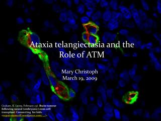
Ataxia telangiectasia • Ataxia telangiectasia (A-T) (Boder-Sedgwick syndrome or Louis–Bar syndrome is a rare, neurodegenerative, inherited disease that affects many parts of the body and causes severe disability. Ataxia refers to poor coordination and telangiectasia to small dilated blood vessels, both of which are hallmarks of the disease. A child who has inherited A-T will display nervous system abnormalities by age 2, and will then progressively lose muscle control.
Genetics: • Ataxia telangiectasia is caused by sequence disruption in the gene ATM (Ataxia telangiectasia mutated). It is an autosomal (relating to one of the 23 chromosomes other than the sex chromosome) recessive disease which means that it will not affect the body unless it has twin copies of the recessive genetic anomaly. Therefore if both the parents carry one copy of the gene, they themselves won't be affected by it, but they will be carriers.
A-T usually runs in families. The mode of inheritance is autosomal recessive, so in a family with two parents who are carriers of the A-T,there is 1 chance in 4 that each child born to the parents will have the disorder. Prenatal diagnosis can be carried out in most families, but this is complex and must be arranged before conception.
Pathophysiology • AT is caused by a defect in the gene responsible for recognizing and correcting errors in duplicating DNA when cells divide. The gene normally repairs double-stranded DNA breaks. • The gene, ataxia-telangiectasia mutated (ATM), discovered in 1995, is on chromosome 11 (11q 22-23). • Normally, when a cell tries to duplicate damaged DNA, it identifies the damage at several checkpoints in the cell division cycle. It tries to repair the damage, and, if it can't repair the damage, it commits suicide through programmed cell death (apoptosis). The ATM gene plays a critical role in this process. It mobilizes several other genes try to repair the DNA damage or destroy the cell if they can't repair it. These downstream genes include tumor suppressor proteins p53 and BRCA1, checkpoint kinase CHK2, checkpoint proteins RAD17 and RAD9, and DNA repair protein NBS1.
In A-T, the pathways that control these processes are defective. This allows cells with damaged DNA to reproduce, resulting in chromosome instability, abnormalities in genetic recombination, and an absence of programmed cell death. ATM patients are particularly sensitive to X-rays, because X-rays induce double-stranded DNA breaks, which they are unable to repair. They are also particularly susceptible to cancers that result from double-stranded DNA breaks. For example, female ATM patients have a two-fold higher risk of developing breast cancer, often before age 50.
Mutations in the ATM gene are thought to come in two types: • Null mutations cause complete loss of function of the protein, and are therefore inherited in a recessive manner and cause A-T. • Missense mutations, which produce stable, full sized protein with reduced function, e.g., substitutions, short in-frame insertions and deletions etc. These mutations act by dominantly interfering with the normal copy of the protein.
The majority of A-T sufferers, 65-70%, have truncating mutations, with exon skipping mutations being particularly common. This results in very low or undetectable levels of ATM protein. Missense mutations are the most common type of mutation found in carriers with breast cancer. Individuals with two missense mutations are believed to have a milder form of AT, which may account for cases of attenuated A-T. Therefore it is thought that "subtle constitutional alterations of ATM may impart an increased risk of developing breast cancer and therefore act as a low penetrance, high prevalence gene in the general population" (Maillet et al. 2002).
ATM Carriers • Carriers of ATM missense mutations are believed to have a 60% penetrance by age 70 and a risk of breast cancer 16 times higher that of the normal population, with a 5-8 fold increased risk of cancer. On average carriers die 7–8 years earlier than the normal population, often from heart disease. Some papers state a lifetime risk for people with both null and missense mutations of 10-38%, which is still a hundredfold increase from population risk. • Individuals with a single ATM mutation are also at a higher risk from lung, gastric and lymphoid tumours, as well as breast cancer. S707P is known to be particularly common in breast cancer patients and F1463S is known to be associated with Hodgkin’s lymphoma. If pulmonary infections could be completely eradicated A-T is consistent with survival into the 5th or 6th decade.
The symptoms: • At first, infants with A-T appear healthy. By age 2, however, parents notice increased clumsiness and balance problems. As symptoms become progressively worse, speech becomes slurred and difficult. Between ages 2 to 8, the telangiectases - tiny, red “spider” veins - appear on the cheeks, ears, and in the eyes. By age 10 to 12, children with A-T lose muscle control.
Other symptoms • can vary, but include immune system deficiencies, low immunoglobulin concentrations. • missing or abnormally developed thymus gland, • retarded growth, • diabetes • prematurely graying hair, • and difficulty swallowing.
Ocular apraxia (difficulty following objects across visual field) • Telangiectasias of the eyes and skin • Chromosomal instability • Hyper-sensitivity to ionizing radiation • Increased incidence of malignancies (primarily hematologic). • Raised alpha-fetoprotein levels. • Absent thymic shadow on X-ray. • Ovarian dysgenesis.
Diagnosis • Diagnosis is usually achieved clinically by examination and identification of both ataxia and oculo-telangiectasia or skin telangiectasia. • This is then followed by laboratory tests for serum AFP level, • the response of white blood cells to X-rays and measurement of the level of ATM protein Sufferers may also have a low lymphocyte count and other immunological abnormalities.
This can then be followed by cytogenetic and molecular testing to confirm the diagnosis. Molecular diagnosis of A-T can be carried out by sequencing all 66 exon of the gene or by linkage if there is a significant family history. • MRI and CT scans may show signs of cerebellar atrophy. (MRI is the preferred method, as patients should limit exposure to any radiological diagnostic tests that use ionizing radiation)
Protein functionality testing is also available. • However A-T testing is usually carried out cytogenetically as specific breakpoints and cytogenetic instability are major characteristic features of the disorder. This must be carried out on lymphocytes. 10% of patients with A-T show balanced translocations, 2/3rds of which involve the immunoglobulin genes on chromosomes 7 and 14. Some patients show expansions in their immunoglobulin genes, which can expand during mitosis resulting in prolymphocyte leukaemia.
Antenatal diagnosis can be carried out using linkage and microsatellite markers. However, direct gene analysis between known sufferers and the foetus is more common. • Histopathologic studies of the brain of an individual with ataxia-telangiectasia have revealed loss of Purkinji cells, granular cells, and basket cells of cerebellar cortex.
Differential Diagnosis • Other Problems to Be Considered - Hartnup disease - Cockayne syndrome - De Sanctis-Cocchione syndrome - Friedreich ataxia - Rendu-Osler-Weber disease
Management • Treatment is symptomatic and supportive. • Physical and occupational therapy may help maintain flexibility. • Speech therapy may also be needed. • Gamma-globulin injections may be given to help supplement a weakened immune system. • High-dose vitamin regimens may also be used. • Antibiotics are used to treat infections. • Some physicians recommend low doses of chemotherapy to reduce the risk of cancer but this is controversial.
It is also recommended that heterozygote family members are regularly monitored for cancers. • Recently deferoxamine was shown to increase the stability of A-T cells and may prove to be an effective treatment for the disorder.
People with A-T have an increased incidence (probably 1% risk per year) of tumours, particularly lymphomas and leukaemia, but due to sufferers' hyper-sensitivity to ionising radiation, radiotherapy and chemotherapy are rarely used.
Prognosis • Those with A-T usually die in their teens or early 20s although some individuals have been known to live to over 40. Mortality is mainly due to the compromised immune system, which causes recurrent respiratory infections, predisposition to cancer, and a high rate of pulmonary problems.
Epidemiology • This disease has had cases all over the world in all races and it's probable that 1 person in 100,000 will be affected by it. Also it has an equal probability of striking in males as well as females. • The incidence of A-T in Caucasians is about 3 per million so the disorder is very rare, with probably fewer than 200 affected people in the UK.
Sex • Males and females are affected equally. • Age • The age of patients with A-T at the time of presentation is 2.5-7 years.
Follow-up • Further Outpatient Care Respiratory infections should be monitored. Physical therapy is indicated. • Prevention The gene responsible for A-T, the ATM gene, was discovered in 1995. This gene makes a protien that activates a number of the protiens that control cell cycle, DNA repair, and cell death. There is ongoing preclinical and early clinical research on gene therapy to treat A-T and other disease.
Complications • Complications may include the following: • Recurrent pulmonary infections. • Progressive ataxia results in the patient being wheelchair ridden. • Death. Patient Education • Children with A-T should have psychologic counseling ase the age because of the great disparity between chronological age and mental age in tests involving visual motor coordination.
References: • http://www.neuroskills.com/brain.shtml • http://www.mayoclinic.com/health/ataxia/DS00910 • http://www.umm.edu/imagepages/18008.htm • http://en.wikipedia.org/wiki/Ataxia_telangiectasia