Innovative Lateral Force Microscopy Sheds Light on Pentacene Microstructure on SiO2
10 likes | 125 Vues
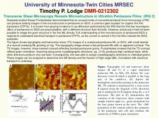
Graduate student Kanan Puntambekar has pioneered a novel method using conventional lateral force microscopy (LFM) to capture detailed images of the microstructure of pentacene on SiO2, a common insulator in organic thin film transistors (OTFTs). This ground-breaking approach reveals the grain structure of polycrystalline pentacene monolayers, paving the way for a deeper understanding of electrical transport properties in OTFTs. High contrast transverse shear images showcase the unique grain orientations and can help determine grain boundary densities and their correlation with electrical performance.

Innovative Lateral Force Microscopy Sheds Light on Pentacene Microstructure on SiO2
E N D
Presentation Transcript
Graduate student Kanan Puntambekar demonstrated that an unusual mode of conventional lateral force microscopy (LFM) can produce striking images of the microstructure in pentacene on SiO2, a common gate insulator for organic thin film transistors (OTFTs). It is known from grazing incidence X-ray diffraction performed by the IRG that the first few monolayers (MLs) of pentacene on SiO2 are polycrystalline, with a different structure than the bulk. However, previously it had not been possible to image the grain structure in the first ML directly. Full understanding of the microstructure of pentacene/SiO2 is required to understand electrical transport in pentacene OTFTs, as the current is carried in the first MLs nearest the SiO2 substrate. The figure shows topography and transverse shear (TS) images of a coalesced pentacene ML on SiO2, with small islands of a second overlying ML growing on top. The topography image shows a flat pentacene ML with no apparent contrast. The TS images, however, show marked contrast reflecting faceted pentacene grains. Puntambekar showed that the TS contrast reflects grain orientation. Scanning along some crystallographic directions, eg., [110], produces high cantilever twist, while other directions produce very low twist. Consequently, she was able to assign crystallographic directions to individual grains. These images can be analyzed to determine the GB density and the fraction of high angle GBs. Correlation with electrical transport is underway. University of Minnesota-Twin Cities MRSECTimothy P. Lodge DMR-0212302Transverse Shear Microscopy Reveals Microstructure in Ultrathin Pentacene Films (IRG 2) Figure. Topography (A) and transverse shear images (B and C) of a vapor deposited pentacene ML on SiO2. (D) defines the scan direction vector S which is parallel to the long axis of the cantilever. (E) depicts the herringbone molecular packing in the pentacene ML. TS contrast is maximized for a scan vector S aligned along the diagonal <110> directions and is minimized for S aligned along the a or b directions. The plot in (F) demonstrates the variation of the transverse shear contrast with sample rotation angle (i.e., grain orientation) for the four grains shown in the inset. The ~180o periodicity of the contrast reflects the symmetry of the pentacene unit cell and confirms the sensitivity of TSM to grain orientation.