Download
1 / 58
710 likes | 1.5k Vues
Echocardiography in Pulmonary Embolism. Gregory Piazza, M.D. Beth Israel Deaconess Medical Center January 26, 2005. Objectives. To present a brief overview of the epidemiology, pathophysiology, diagnosis, and management of acute pulmonary embolism (PE).

E N D
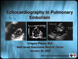
Echocardiography in Pulmonary Embolism Gregory Piazza, M.D. Beth Israel Deaconess Medical Center January 26, 2005 Beth Israel Deaconess Medical Center
Objectives • To present a brief overview of the epidemiology, pathophysiology, diagnosis, and management of acute pulmonary embolism (PE). • To review the role of echocardiography in the diagnosis of PE. • To highlight the role of echocardiography in risk stratification of patients with PE. Beth Israel Deaconess Medical Center
Epidemiology • The incidence of PE in the U.S. is approximately 1 per 1000 per year. • Only 1 out of every 3 cases of venous thromboembolism (VTE), including DVT and PE, is diagnosed. • With approximately 450,000 cases detected per year, a staggering 900,000 VTE cases may go undiagnosed annually. Lancet 1999;353:1386-1389 Lancet 2004;363:1295-1305 Beth Israel Deaconess Medical Center
Epidemiology • In the Olmsted County registry, 30-day mortality after PE or DVT has been reported as high as 28%. • The International Cooperative Pulmonary Embolism Registry (ICOPER) estimates a 3-month mortality of 17.4%. • These data suggest PE is possibly as deadly as acute myocardial infarction. Arch Intern Med 1999;159:445-453 Circulation 2003;108:2726-2729 Beth Israel Deaconess Medical Center
Pathophysiology • The most common sources of PE are the deep veins of the lower extremities and pelvis. • Thrombi dislodge from these veins and embolize to the pulmonary arterial tree where they trigger pathophysiologic changes in hemodynamics and gas exchange. • The size of the embolus, underlying cardiopulmonary status, and neurohumoral adaptations determine the hemodynamic response to PE. Circulation 2003;108:2726-2729 Beth Israel Deaconess Medical Center
Pathophysiology www.benlovejoy.com/ pulmonary_embolism_main.html Beth Israel Deaconess Medical Center
Pathophysiology • Physical obstruction, release of vasoconstrictors, and hypoxia lead to increased pulmonary vascular resistance (PVR) and right ventricular (RV) afterload. • RV pressure overload leads to chamber dilatation and hypokinesis, tricuspid regurgitation, and eventual RV failure. • RV pressure overload also causes interventricular septal flattening during systole and deviation towards the left ventricle (LV) during diastole leading to impaired LV filling. • As RV pressure overload worsens, RV wall stress and ischemia develop secondary to increased myocardial oxygen demand and decreased supply. Am Heart J 1995;130:1276-1282 Beth Israel Deaconess Medical Center
Pulmonary embolism ↑ PA pressure ↑ RV afterload ↑ RV wall tension ↑ RV O2 demand RV dilatation and dysfunction RV ischemia +/- infarction ↓ RV O2 supply ↓ RV cardiac output Septal shift toward the LV ↓ coronary perfusion ↓ LV preload ↓ LV cardiac output Hypotension Pathophysiology Am Heart J 1995;130:1276-1282 Beth Israel Deaconess Medical Center
Spectrum of Disease • A variety of clinical syndromes may be seen: • Normotensive with normal RV function • Normotensive with RV dysfunction (submassive PE) • Cardiogenic shock (massive PE) • Cardiac arrest (massive PE) Beth Israel Deaconess Medical Center
History: Dyspnea (most frequent symptom) Pleuritic chest pain Cough Hemoptysis Syncope Physical: Tachypnea (most frequent sign) Anxious appearance Tachycardia Fever Elevated JVD (most specific sign) Loud P2 Tricuspid regurgitation Paradoxical bradycardia Diagnosis: History and Physical Beth Israel Deaconess Medical Center
Arterial blood gases Electrocardiography Chest X-ray Plasma D-dimer Lower extremity ultrasound Echocardiography (TTE and TEE) Ventilation-perfusion lung scanning Spiral chest CT Magnetic resonance (MR) angiography Contrast pulmonary angiography The Diagnostic Armamentarium Beth Israel Deaconess Medical Center
History and Physical Eval. clinical likelihood Patient in ED Electrocardiogram Chest radiograph Patient already in hospital D-dimer Normal High V/Q if dye allergy or renal insufficiency Chest CT No PE Normal Positive Equivocal Normal Ultrasonography Positive Negative No PE Treat for PE Consider PA-gram No PE Diagnosis: An Integrated Approach Lancet 2004;363:1295-1305 Beth Israel Deaconess Medical Center
Diagnosis: Transthoracic Echocardiography • Transthoracic echocardiography (TTE) is insensitive in the diagnosis of acute PE. • In a prospective study, TTE failed to diagnose 50% of patients with angiographically proven PE. • However, in the appropriate clinical setting, findings of right ventricular pressure overload may help suggest acute PE as a diagnosis. Lancet 2004;363:1295-1305 Am J Med 2001;110:528-535 Beth Israel Deaconess Medical Center
Apical 4- chamber Parasternal short-axis Parasternal long-axis Diagnosis: Transthoracic Echocardiography Am J Respir Crit Care Med 2002;166:1310-1319 Beth Israel Deaconess Medical Center
Echocardiographic Findings In Acute PE • RV dilatation and hypokinesis • Interventricular septal flattening and paradoxical motion • Alteration of transmitral gradients with A wave > or = E wave • Tricuspid regurgitation (TR) • Pulmonary artery (PA) hypertension as estimated by the modified Bernoulli equation • RA dilatation • Loss of respiratory-phasic IVC collapse with inspiration • Patent foramen ovale • RA, RV, or pulmonary artery thrombus Ann Intern Med 2002;136:691-700 Beth Israel Deaconess Medical Center
RV Dilatation • In the apical 4 chamber view, a ratio RVEDA (area) to LVEDA > 0.6 correlates with moderate RV dilatation. • A ratio > or = 1.0 correlates with major RV dilatation. Am J Respir Crit Care Med 2002;166:1310-1319 Beth Israel Deaconess Medical Center
RV Hypokinesis • RV hypokinesis is frequently diagnosed in a qualitative fashion. • Quantitative methods, such as RV fractional area contraction, have not proven more accurate. • McConnell et al. noted a specific qualitative finding in patients with RV dysfunction and acute PE compared to patients with other causes of RV failure. • The McConnell sign is noted when RV free-wall hypokinesis in observed in the setting of relatively normal RV apical contraction. Am J Respir Crit Care Med 2002;166:1310-1319 Ann Intern Med 2002;136:691-700 Am J Cardiol 1996;78:469-473 Beth Israel Deaconess Medical Center
RV Hypokinesis • The RV free-wall endocardium was traced in the apical 4-chamber view from base to apex at end-systole and end-diastole. • Tracings from patients with RV dysfunction from acute PE were compared to those with RV dysfunction from pulmonary arterial hypertension. • For PE, the McConnell sign had a sensitivity of 77%, specificity of 94%, PPV of 71%, and NPV of 96%. Am J Cardiol 1996;78:469-473 Beth Israel Deaconess Medical Center
RV Hypokinesis Courtesy of A. Rosen Beth Israel Deaconess Medical Center
Diastole Systole Interventricular septal flattening and paradoxical motion • Right ventricular pressure overload leads to deviation of the interventricular septum towards the LV in diastole. • Interventricular septal flattening is seen during systole creating a so-called D-shaped LV. Am J Respir Crit Care Med 2002;166:1310-1319 Ann Intern Med 2002;136:691-700 J Am Coll Cardiol 1987;10:1201-1206 Beth Israel Deaconess Medical Center
Normal (diastole) Acute PE (diastole) Interventricular septal flattening and paradoxical motion Am J Respir Crit Care Med 2002;166:1310-1319 Beth Israel Deaconess Medical Center
Normal PE Alteration of Transmitral Gradients • In the setting of pericardial constraint, interventricular septal motion towards the LV during diastole leads to impaired LV filling. • Diastolic impairment leads to an A wave that is > or = to the E wave, signifying increased dependence on atrial contraction for LV filling. Am J Respir Crit Care Med 2002;166:1310-1319 Beth Israel Deaconess Medical Center
Alteration of Transmitral Gradients Courtesy of A. Rosen Beth Israel Deaconess Medical Center
RV RA LA Tricuspid Regurgitation • RV pressure overload frequently results in tricuspid regurgitation detected on color flow and Doppler. Ann Intern Med 2002;136:691-700 www.geocities.com/SouthBeach/ Cove/2045/echo55.htm Beth Israel Deaconess Medical Center
Pulmonary Hypertension • PA systolic pressure is estimated by using the modified Bernoulli equation: P = 4V2 where P = peak pressure gradient V = peak velocity of the TR jet • Estimated RA pressure is added to the gradient to approximate PA systolic pressure. Courtesy of A. Rosen Am J Respir Crit Care Med 2002;166:1310-1319 Ann Intern Med 2002;136:691-700 Beth Israel Deaconess Medical Center
Thrombus In The Right Main PA Ann Intern Med 2002;136:691-700 Beth Israel Deaconess Medical Center
Diagnosis: Transesophageal Echocardiography • Transesophageal echocardiography (TEE) can diagnose PE by direct visualization of the proximal pulmonary arteries. • Because the left main bronchus obstructs the view of the middle portion of the left pulmonary artery, PE is more difficult to detect in the left PA. • TEE may play a unique role in the diagnosis of PE in patients with unexplained cardiac arrest (especially pulseless electrical activity). Ann Intern Med 2002;136:691-700 Beth Israel Deaconess Medical Center
Long-axis transesophageal Short-axis transgastric Oblique transgastric Diagnosis: Transesophageal Echocardiography Am J Respir Crit Care Med 2002;166:1310-1319 Beth Israel Deaconess Medical Center
Diagnosis: Transesophageal Echocardiography Ann Intern Med 2002;136:691-700 Beth Israel Deaconess Medical Center
Case Study • A 67 year old male with history of CAD, HTN, and prostate cancer presents with acute onset dyspnea and dull chest pressure. • On exam, he is tachycardic, tachypneic, hypoxic, but normotensive. He has elevated neck veins and new lower extremity edema. • His EKG reveals sinus tachycardia. • His chest X-ray is read as “no pneumonia, no CHF.” • Because of high clinical suspicion for PE, he undergoes chest CT. Beth Israel Deaconess Medical Center
Case Study Beth Israel Deaconess Medical Center
Case Study • The patient is started on a weight-based protocol of intravenous unfractionated heparin and admitted to a telemetry floor. • That evening, the patient’s roommate calls the nurses station to report that the patient has “slumped over in his chair.” • The patient is found unresponsive and a code is called. • The patient is found to be in pulseless electrical activity (PEA) and expires after resuscitative efforts are unsuccessful. Beth Israel Deaconess Medical Center
Risk Stratification Risk Stratification Tools: • History and physical • Clinical prognostic scores • Cardiac biomarkers including cardiac troponin and brain-type natriuretic peptide (BNP) • Chest CT • Echocardiography Beth Israel Deaconess Medical Center
History and Physical • ICOPER reported several independent clinical predictors of increased mortality at 3 months. Beth Israel Deaconess Medical Center Lancet 1999;353:1386-9
Cardiac Biomarkers • Cardiac troponins and BNP have been extensively studied in the evaluation of patients with acute PE. • Cardiac troponins and BNP accurately identify low-risk PE patients with negative predictive values for in-hospital death ranging from 97 to 100%. • Patients presenting with acute PE and elevated cardiac biomarkers should undergo transthoracic echocardiography to assess RV function. • In patients with acute PE and normal levels of cardiac biomarkers, echocardiography is not routinely required as RV function will most often be normal. Circulation 2003;108:2191-2194 Beth Israel Deaconess Medical Center
Cardiac Biomarkers Accuracy of cardiac biomarkers for the prediction of in-hospital death in acute pulmonary embolism. Beth Israel Deaconess Medical Center Circulation 2003;108:2191-2194
↑ PVR ↑ RV pressure RV micro-infarction ↑ RV shear stress Myofibril degradation ↑ Natriuretic peptide mRNA ↑ Troponins ↑ BNP Cardiac Biomarkers Beth Israel Deaconess Medical Center Circulation 2003;108:2191-2194
Chest CT Scan • Although chest CT is used primarily for the diagnosis of PE, RV dilatation may also be observed. • In a study of 431 patients with acute PE diagnosed by chest CT, multiplanar reformats of axial CT data into CT 4-chamber views were performed. • Right and left ventricular dimensions (RVD and LVD) were measured. RV enlargement was defined as RVD /LVD > 0.9. • RV enlargement predicted 30-day death (hazard ratio, 5.17, p = 0.005) after adjusting for pneumonia, cancer, chronic lung disease, and age. Beth Israel Deaconess Medical Center Circulation 2004;110:3276-3280
Chest CT Scan Beth Israel Deaconess Medical Center Circulation 2004;109:2401-2404
Echocardiography • RV dysfunction on echocardiography has been reliably established as a predictor of adverse outcomes in PE. • The most commonly accepted quantitative standards are: 1. RV to LV end-diastolic diameter ratio > 1 in the apical 4-chamber view 2. RV end-diastolic diameter > 30 mm 3. Paradoxical interventricular septal systolic motion Ann Intern Med 2002;136:691-700 Beth Israel Deaconess Medical Center
Echocardiography • At the Karolinska Institute in Sweden, 126 consecutive patients with PE were examined with TTE on the day of diagnosis. • After multivariate analysis, RV dysfunction emerged as the most powerful predictor of in-hospital death. • A 6-fold increase in relative risk was noted in the patients with RV dysfunction compared to those with normal RV function. Am Heart J 1997;134:479-487 Beth Israel Deaconess Medical Center
Echocardiography • In a cohort of 209 consecutive patients with PE, 31% presented with a combination of normal blood pressure and echocardiographic evidence of RV dysfunction. • Of these patients, 10% developed cardiogenic shock within 25 hours and 5% died in hospital. • None of the patients with normal RV function died from PE. Circulation 2000;101:2817-2822 Beth Israel Deaconess Medical Center
Echocardiography • In ICOPER, 90-day mortality rate was increased in patients with RV dysfunction. • After multiple regression analysis, RV dysfunction was found to be an independent predictor of death at 90 days. Lancet 1999;353:1386-1389 Beth Israel Deaconess Medical Center
No shock Shock BNP ↓ Troponin ↓ BNP ↑ Troponin ↑ Echocardiography No RV dysfunction RV dysfunction Anticoagulation alone Consider thrombolysis or embolectomy Risk Stratification Algorithm Beth Israel Deaconess Medical Center Circulation 2003;108:2191-2194
Primary therapy: Thrombolysis Open surgical embolectomy Catheter-assisted embolectomy Secondary therapy: IV unfractionated heparin Low-molecular weight heparin (LMWH) Fondaparinux Warfarin IVC filter Management Beth Israel Deaconess Medical Center
Management • In patients with massive PE, primary therapy with thrombolytics is considered a lifesaving intervention. • Surgical or catheter-assisted embolectomy may be considered for massive PE if thrombolysis is contraindicated. • For submassive PE, thrombolysis remains controversial as no mortality benefit has been shown in this patient population. • However, MAPPET-3 demonstrated a reduction in need for escalation of therapy in patients receiving up-front t-PA (alteplase) for submassive PE. • Normotensive patients with normal RV function are considered low-risk and receive standard anticoagulation. J Thromb Thrombolysis 1995;2:227-229 N Engl J Med 2002;347:1143-1150 Beth Israel Deaconess Medical Center
Thrombolysis in PE: Pre-Lytics *Following echo loops are courtesy of A. Kothavale Beth Israel Deaconess Medical Center


