Download
1 / 36
360 likes | 364 Vues
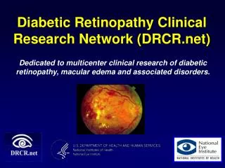
The Diabetic Retinopathy Clinical Research Network. An Observational Study of the Development of Diabetic Macular Edema Following Panretinal (Scatter) Photocoagulation (PRP) Given in 1 or 4 Sittings Sponsored by the National Eye Institute,

E N D
The Diabetic Retinopathy Clinical Research Network An Observational Study of the Development of Diabetic Macular Edema Following Panretinal (Scatter) Photocoagulation (PRP) Given in 1 or 4 Sittings Sponsored by the National Eye Institute, National Institutes of Health, U.S. Department of Health and Human Services.
Background: Panretinal Photocoagulation (PRP) for PDR • PRP is the standard treatment of PDR • Usually performed over 1 or more sittings (ETDRS 2 or more sittings) • 2004 survey of DRCR.net investigators: one-quarter routinely use one sitting for PRP
PRP and Macular Edema • PRP associated complications include macular edema • ETDRS - 18% developed macular edema 4 months after full PRP (1200 – 1600 spots) • Theories: • Oncotic fluid accumulation related to tissue destruction • PRP induced inflammation leading to cytokine release and increased permeability of the retinal capillaries
What are Some Pros and Cons of Delivering PRP in 1 Sitting? • Pros • Convenience • Completion before complications occur [e.g., vitreous hemorrhage (VH)] • Cons (theoretical): • May cause macular edema with VA loss • Greater patient discomfort • Need for retrobulbar or peribulbar anesthetic • Higher risk of acute pressure rise, angle closure, or choroidal effusion
What are Some Pros and Cons of Delivering PRP in More Than One Sitting? • Pros (theoretical): • Allows edema and associated VA loss to subside before next PRP sitting • Cons: • Inconvenient • Potential for failure to complete intended treatment plan (VH)
Primary Study Objective • Determine the incidence of new macular edema on OCT, following PRP in diabetic eyes without center edema at 34 weeks • To compare the incidence of macular edema with a 1-sitting versus 4-sitting regimen
Non-Randomized Study: Rational • Investigators who complete PRP in one sitting were unwilling to perform PRP in multiple sittings and vice versa • Therefore, prospective nonrandomized study conducted
Study Design Prospective, Non-Randomized Study Major Eligibility Criteria Assessed: >18 years old Type 1 or type 2 diabetes Early-PDR or Severe-NPDR (high risk PDR not eligible) Retinal thickness on OCT < 300 microns in the central subfield Visual Acuity letter score > 73 (20/32 or better) No clinical DME requiring treatment Investigator declared, prior to study initiation, if PRP planned for 1 or 4 sittings for all subjects participating in protocol 1 Sitting 4 Sittings
PRP Treatment Regiment • 1200-1600 total burns (ETDRS parameters) • Retrobulbar/peribulbar anesthesia optional • Slit lamp delivery systems 1 Sitting and 4 Sitting • Each session 4 weeks apart (+4 days) • 300 burns in each of first two sittings • Investigator discretion for # of burns in 3rd and 4th sittings • Total of 4 sittings between 1200 and 1600 burns 4 Sittings
PRP Burn Characteristics Laser Parameters:
Outcome Assessment • Visual acuity measured at 3 meters with the Electronic-ETDRS Visual Acuity (EVA) device • Central Subfield Thickness Using Zeiss Stratus OCT 3 Day 4 Week 17 Week • Central Subfield Thickness • Primary Outcome • E-ETDRS Visual Acuity • 3-Field Fundus Photographs 34 Week
Study Enrollment 155 subjects enrolled at 27 sites 1 sitting 84 subjects 4 sittings 71 subjects 34-week Visit Completion (Primary Outcome) 1 sitting 88% 4 sittings 82% 13
Baseline Characteristics Women: 48% 44% Median Age: 56 yrs 54 yrs Race: • White 64% 46% • African-American 24% 28% • Hispanic or Latino 11% 18% • Asian 0 6% • More than one race 1% 1% 4 Sitting 1 Sitting
Baseline Characteristics 4 Sitting 1 Sitting Diabetes Type • Type 1 19% 25% • Type 2 81% 75% Median Duration of Diab. 18 yrs 20 yrs Median HbA1c (%)* 7.7 8.2 *HbA1c: Missing 7 in one-sitting group, 13 in 4 sitting group
Baseline Characteristics 4 Sitting 1 Sitting Prior Focal Macular Laser 14% 11% Median E-ETDRS Letter Score 85 83 (Snellen equivalent) 20/20 20/20 Median OCT CSF Thickness 207µ 198µ CSF 250 – 299 Microns 3 (4%) 1 (1%) Median OCT Retinal Volume 6.9 mm3 7.0 mm3
Baseline Retinopathy Severity 4 Sitting 1 Sitting N 81 66 Mild NPDR 6% 0 Moderate NPDR 9% 3% Moderately Severe NPDR 30% 38% Severe NPDR 6% 8% Mild PDR 23% 15% Moderate PDR 14% 15% High Risk PDR 11% 18% Cannot Grade 1% 3%
Scatter Treatment 4 Sitting 1 Sitting N 84 71 Median # of Burns Median Average Power Retrobulbar Injection Incomplete Treatment Additional PRP 4 Sitting 1 Sitting 1274 280mW 46% 0% 7% 1260* 250mW 14%** 15% 0% * Total over all 4 sittings. **At least once
Central Subfield Thickness 1-sitting 4-sitting P value Baseline 207µ 198µ Change from baseline - median microns • 3d +9 +5 0.01 • 4wks +13 +5 0.003 • 17wks +14 +15 0.08 • 34wks +14 +22 0.06
Treatment of DME • One eye in each group was treated for DME prior to 34 weeks
E-ETDRS Visual Acuity 1 Sitting 4 Sitting P Value Baseline 85 83 Change from baseline • 3d−3 −1 0.005 • 4wks−1 −1 0.37 • 17wks−1 −1 0.66 • 34wks 0 −2 0.006 Mean Letter Score 24
E-ETDRS Visual Acuity: 3 Day N=82 N=66 2%
E-ETDRS Visual Acuity: 4 Week N=78 N=67
E-ETDRS Visual Acuity: 17 Week N=77 N=63
E-ETDRS Visual Acuity: 34 Week N=72 N=56 17% 2%
Vitreous Hemorrhage • A vitreous hemorrhage reducing visual acuity by 10 or more letters occurred in 2 eyes in each group.
Summary • Treatment groups balanced on baseline factors • Number of total burns similar between treatment groups • Retrobulbar anesthesia more commonly used in 1-sitting group
Summary • CSF Thickness Remained Normal in Most Eyes • 3 Days – • CSF Greater in 1 than 4 Sitting Group • 34 Weeks – • CSF Greater in 4 than 1 Sitting Group • Differences Unlikely to be Clinically Relevant • VA and OCT Results Consistent
Conclusion • Eyes Without Edema • Single Sitting PRP – • Safe • Little Change in VA or Edema • 1 Sitting vs. 4 Sitting PRP – • Clinically Meaningful Differences Unlikely • VA and OCT Changes Similar
Study Limitations 1 Sitting vs. 4 Sitting PRP • Small Sample Size • Only Large Differences Ruled Out • Non – Randomized Study Design • Uncontrolled Confounding Possible
Conclusion 1 Sitting vs. 4 Sitting PRP Results Apply Only to Eyes Without Edema Impact if 1 & 4 Sittings Clinically Equivalent One Sitting Benefits and Risks Increased Patient Convenience Decreased Patient Cost – Travel & Productivity Possible Risk of Retrobulbar Anesthesia Unknown if results apply to eyes with DME 35
Thank You • Subjects who volunteered to participate in this trial • 27 clinical study sites investigators, coordinators, and other staff • DRCR Network Data and Safety Monitoring Committee • DRCR Network General Steering Committee • Other DRCR Network investigators and staff • Sponsor: National Eye Institute of the National Institutes of Health (U.S. Department of Health and Human Services For further information and all DRCR Network financial disclosures, go to www.drcr.net 36