Download
1 / 1
10 likes | 166 Vues
UNIVERSIDADE FEDERAL DA BAHIA INSTITUTO DE CIÊNCIAS DA SAÚDE LABORATÓRIO DE IMUNOLOGIA E BIOLOGIA MOLECULAR COMPARATIVE STUDY OF DIFFERENT PROTEIC ANTIGENIC EXTRACTS OF C orynebacterium pseudotuberculosis

E N D
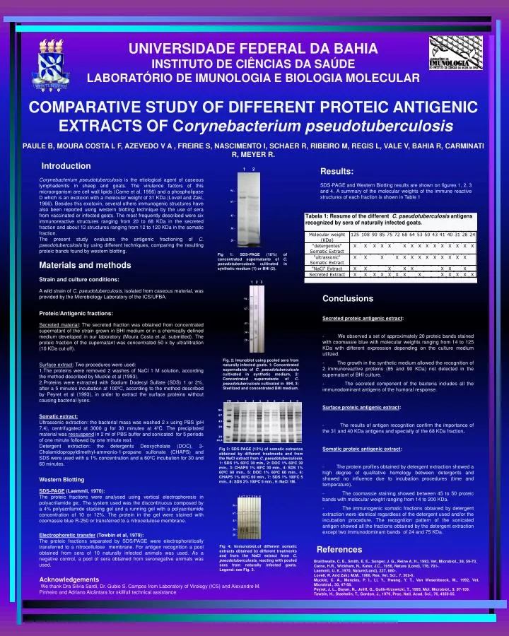
UNIVERSIDADE FEDERAL DA BAHIA INSTITUTO DE CIÊNCIAS DA SAÚDE LABORATÓRIO DE IMUNOLOGIA E BIOLOGIA MOLECULAR COMPARATIVE STUDY OF DIFFERENT PROTEIC ANTIGENIC EXTRACTS OF Corynebacterium pseudotuberculosis PAULE B, MOURA COSTA L F, AZEVEDO V A , FREIRE S, NASCIMENTO I, SCHAER R, RIBEIRO M, REGIS L, VALE V, BAHIA R, CARMINATI R, MEYER R. 1 2 • Introduction • Corynebacterium pseudotuberculosis is the etiological agent of caseous lymphadenitis in sheep and goats. The virulence factors of this microorganism are cell wall lipids (Carne et al, 1956) and a phospholipase D which is an exotoxin with a molecular weight of 31 KDa (Lovell and Zaki, 1966). Besides this exotoxin, several others immunogenic structures have also been reported using western blotting technique by the use of sera from vaccinated or infected goats. The most frequently described were six immunoreactive structures ranging from 20 to 68 KDa in the secreted fraction and about 12 structures ranging from 12 to 120 KDa in the somatic fraction. • The present study evaluates the antigenic fractioning of C. pseudotuberculosis by using different techniques, comparing the resulting proteic bands found by western blotting. • Materials and methods • Strain and culture conditions: • A wild strain of C. pseudotuberculosis, isolated from caseous material, was provided by the Microbiology Laboratory of the ICS/UFBA. • Proteic/Antigenic fractions: • Secreted material: The secreted fraction was obtained from concentrated supernatant of the strain grown in BHI medium or in a chemically defined medium developed in our laboratory (Moura Costa et al, submitted). The proteic fraction of the supernatant was concentrated 50 x by ultrafiltration (10 KDa cut off). • Surface extract: Two procedures were used: • The proteins were removed 2 washes of NaCl 1 M solution, according the method described by Muckle et al (1993). • Proteins were extracted with Sodium Dodecyl Sulfate (SDS) 1 or 2%, after a 5 minutes incubation at 100ºC, according to the method described by Peyret et al (1993), in order to extract the surface proteins without causing bacterial lyses. • Somatic extract: • Ultrassonic extraction: the bacterial mass was washed 2 x using PBS (pH 7,4), centrifugated at 3000 g for 30 minutes at 4ºC. The precipitated material was ressuspend in 2 ml of PBS buffer and sonicated for 5 periods of one minute followed by one minute rest. • Detergent extraction: the detergents Deoxycholate (DOC), 3-Cholamidopropyldimethyl-ammonio-1-propane sulfonate (CHAPS) and SDS were used with a 1% concentration and a 60ºC incubation for 30 and 60 minutes. • Western Blotting • SDS-PAGE (Laemmli, 1970): • The proteic fractions were analysed using vertical electrophoresis in polyacrilamide ge;. The system used was the discontinuous composed by a 4% polyacrilamide stacking gel and a running gel with a polyacrilamide concentration of 10 or 12%. The protein in the gel were stained with coomassie blue R-250 or transferred to a nitrocellulose membrane. • Electrophoretic transfer (Towbin et al, 1979): • The proteic fractions separated by SDS/PAGE were electrophoretically transferred to a nitrocellulose membrane. For antigen recognition a pool obtained from sera of 10 naturally infected animals was used. As a negative control, a pool of sera obtained from seronegative animals was used. Results: SDS-PAGE and Western Blotting results are shown on figures 1, 2, 3 and 4. A summary of the molecular weights of the immune reactive structures of each fraction is shown in Table 1 94 - 67 - 43 - Fig 1: SDS-PAGE (10%) of concentrated supernatante of C. pseudotuberculosis cultivated in synthetic medium (1) or BHI (2). 30 - 20 - 1 2 3 Conclusions Secreted proteic antigenic extract: - We observed a set of approximately 20 proteic bands stained with coomassie blue with molecular weights ranging from 14 to 125 KDa with different expression depending on the culture medium utilized. - The growth in the synthetic medium allowed the recognition of 2 immunoreactive proteins (85 and 90 KDa) not detected in the supernatant of BHI culture. - The secreted component of the bacteria includes all the immunodominant antigens of the humoral response. Surface proteic antigenic extract: - The results of antigen recognition confirm the importance of the 31 and 40 KDa antigens and specially of the 68 KDa fraction. Somatic proteic antigenic extract: - The protein profiles obtained by detergent extraction showed a high degree of qualitative homology between detergents and showed no influence due to incubation procedures (time and temperature). - The coomassie staining showed between 45 to 50 proteic bands with molecular weight ranging from 14 to 200 KDa. - The immunogenic somatic fractions obtained by detergent extraction were identical regardless of the detergent used and/or the incubation procedure. The recognition pattern of the sonicated antigen showed all the fractions obtained by the detergent extraction except two immunodominant bands of 24 and 75 KDa. 94 - 67 - 43 - 30 - 20 - Fig. 2: Imunoblot using pooled sero from naturally infected goats. 1: Concentrated supernatante ofC. pseudotuberculosis cultivated in synthetic medium, 2: Concentrated supernatante ofC. pseudotuberculosis cultivated in BHI, 3: Sterilized and concentrated BHI medium. PM 1 2 3 4 5 6 7 8 9 94 - 67 - 43 - 30 - 20 - 14,4 - Fig 3: SDS-PAGE (12%) of somatic extractos obtained by different treatments and from the NaCl extract from C. pseudotuberculosis. 1: SDS 1% 60ºC 30 min., 2: DOC 1% 60ºC 30 min., 3: CHAPS 1% 60ºC 30 min., 4: SDS 1% 60ºC 60 min., 5: DOC 1% 60ºC 60 min., 6: CHAPS 1% 60ºC 60 min., 7: SDS 1% 100ºC 5 min., 8: SDS 2% 100ºC 5 min., 9: NaCl 1M. 1 4 7 8 2 5 3 6 9 94 - 67 - 43 - 30 - 20 - Fig 4: Immunoblot.of different somatic extracts obtained by different treatments and from the NaCl extract from C. pseudotuberculosis, reacting with pooled sera from naturally infected goats. Legend: see Fig. 3. • References • Braithwaite, C. E., Smith, E. E., Songer, J. G., Reine A. H., 1993, Vet. Microbiol., 38, 59-70. • Carne, H.R., Wickham, N., Kater, J.C., 1956, Nature (Lond), 178, 701-. • Laemmli, U. K.,1970, Nature(Lond), 227, 680-. • Lovell, R. And Zaki, M.M., 1966, Res. Vet. Sci., 7, 302-6. • Muckle, C. A., Menzies, P. I., Li, Y., Hwang, Y. T., Van Wesenbeeck, M., 1992, Vet. Microbiol., 30, 47-58. • Peyret, J. L., Bayan, N., Joliff, G., Gulik-Krzywicki, T., 1993, Mol. Microbiol., 9, 97-109. • Towbin, H., Staehelin, T., Gordon, J., 1979, Proc. Natl. Acad. Sci., 76, 4350-55. Acknowledgements We thank Dra Silvia Sardi, Dr. Gubio S. Campos from Laboratory of Virology (ICS) and Alexandre M. Pinheiro and Adriano Alcântara for skillfull technical assistance