Overview of Light Microscopy Techniques and Literature Reference
This document provides a comprehensive overview of light microscopy techniques, focusing on bright-field microscopy and its applications in contrasting modes. Key concepts include the definition of amplitude objects and how staining enhances contrast. Notable literature references include "Optical Physics" by S.G. Lipson, H. Lipson, and D.S. Tannhauser, and "Introduction to Optical Microscopy" by Jerome Mertz. These texts are essential for understanding the principles of optical microscopy, imaging techniques, and contrast enhancement methods in biological specimens.

Overview of Light Microscopy Techniques and Literature Reference
E N D
Presentation Transcript
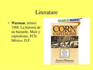
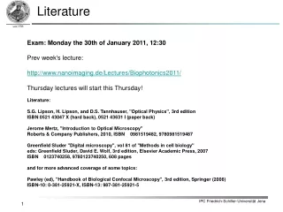
Literature Exam: Monday the 30th of January 2011, 12:30Prev week's lecture:http://www.nanoimaging.de/Lectures/Biophotonics2011/Thursday lectures will start this Thursday!Literature:S.G. Lipson, H. Lipson, and D.S. Tannhauser, "Optical Physics", 3rd editionISBN 0521 43047 X (hard back), 0521 43631 I (paper back)Jerome Mertz, "Introduction to Optical Microscopy"Roberts & Company Publishers, 2010, ISBN 0981519482, 9780981519487Greenfield Sluder "Digital microscopy", vol 81 of "Methods in cell biology"eds: Greenfield Sluder, David E. Wolf, 3rd edition, Elsevier Academic Press, 2007ISBN 0123740258, 9780123740250, 608 pagesand for more advanced coverage of some topics:Pawley (ed), "Handbook of Biological Confocal Microscopy", 3rd edition, Springer (2006)ISBN-10: 0-381-25921-X, ISBN-13: 987-381-25921-5
2. Contrast modes in light microscopy: Bright field • 2.1 Bright field transmission (absorption = imaginary part of refractive index) • An object, keeping the phase of an incoming wave constant and decreasing the amplitude is called amplitude object. • Contrast is A0 –A1,2 • Bright filed microscopy is the most simpleand basic light microscopy method • Sample is illuminated from belowby a light cone • In case there is no sample in the opticalpath a uniform bright image is generated • An amplitude object absorbs light at certain wavelengths and therefore reduces the amplitude of the light passing through the object Amplitude difference Wavelength l Uniform bright field image Bright field image of Moss reeds
2. Contrast modes in light microscopy: Bright field • 2.1 Bright field (absorption = imaginary part of refractive index) • very little absorption: impractical for thin objects • Increase contrast by staining = chemical contrasting: • dyes to mark cell- and tissue structures • Most dyes selectively accumulate within cells (e.g. lipophilic, hydrophilic) • Dyes are often present as ions: • positive charge: cationic or basic dye • anion: anionic or acidic dye • Staining often requires fixation
2. Contrast modes in light microscopy: Bright field • 2.1 Bright field (absorption = imaginary part of refractive index) • Bright field staining: common for histological cross sections: • E.g. hematoxylin and eosin stain:Popular in histology for morphological inspection of biopsy specimen to identify malignant changes • The basic dye hematoxylin colors (blue-purple) basophilic structures which are usually the ones containing nucleic acids: • ribosomes • chromatin-rich cell nucleus • RNA in cytoplasm • Eosin colors (bright pink) eosinophilic structures which are generally composed of protein. hematoxylin and eosin staining of cancer cells
2. Contrast modes in light microscopy: Bright field • 2.1 Bright field (absorption = imaginary part of refractive index) • Gram-staining (crystal violet, alcohol wash, safranin or fuchsin counterstain):Method of differentiating bacterial species into two large groups based on high amount of peptidoglycan in cell walls.: • Gram-positive: bacteria appear after staining dark blue • Gram-negative:crystal violet is washed out. Stained red afterwards by fuchsine or safranin. Bacillus cereus: Gram-positive Pseudomonas aeruginosa: Gram-negative
2. Contrast modes in light microscopy: Bright field Blackboard exercise: Geometric Optics of a Microscope Image Planes and Aperture Planes IPC Friedrich-Schiller-Universität Jena
fTL fTL image plane sample plane back focal plane infinity path : Filters do not hurt The modern microscope: Infinity optics Tube Lens Objective Lens fObj fObj M = fTL / fObj
Telecentric:fTL immersionmedium Meaning of the back focal plane (BFP) Object plane BFP Image plane coverslip Tube lense R a fobj fTL
Optical Aberrations: Spherical Aberration Perfect Lens http://en.wikipedia.org/wiki/Spherical_aberration Real Lens http://en.wikipedia.org/wiki/File:Spherical_aberration_2.svg
Optical Aberrations: Spherical Aberration http://en.wikipedia.org/wiki/File:Spherical-aberration-slice.jpg http://www.olympusmicro.com/primer/java/aberrations/pointspreadaberration/index.html
2. Contrast modes in light microscopy: Bright field Blackboard exercises:Coherent vs. Incoherent imaging The Concept of a Amplitude Spread Function Image Field as a Convolution of Object with ASF The Concept of a Point Spread Function Imaging as a Convolution of Object with PSF
Real Space (PSF) Reciprocal Space (ATF) Lens Cover Glass ky y x kx Focus z kz Oil Intensity in Focus (PSF)
? Fourier Transform ~ ~ ~* I(k) = A(k) A(-k) OTF ATF Epifluorescent PSF I(x) = |A(x)|2 = A(x) · A(x)*
Region of Support kx,y kz Convolution: Drawing with a Brush
! kx,y kz Optical Transfer Function (OTF)
Missing Cone Widefield OTF support
Objective Lense Tube Lense CCD Back FocalPlane 2. Contrast modes in light microscopy: Bright field Scattering / Absorbtion Bright FieldTransmission Dark object on bight backgroundRelative scattering angle and wavelength defines resolutionCondensor AND objective Numerical Aperture matterContrast decreases when resolution increases
kout Kobj kin 2. Contrast modes in light microscopy: Bright field Interference of diffracted lightwith the undiffracted reference (first Born approx.) Range ofDetection Angles "Bragg condition" Holgraphy with plane wave illumination: infinitely little 3D information is acquired!
2. Contrast modes in light microscopy: Bright field 1.? 2.? 3.? 6.? 4.? 5.? http://biology.about.com