Download
1 / 39
410 likes | 729 Vues
Explore the world of microscopes and their types, learn about light properties, compound microscopes, benefits, and applications like comparison microscopy. Discover the advantages of stereoscopic microscopes for specialized viewing needs.

E N D
Microscopy and their types Dr.S.Umamaheswari Assistant Professor M.Sc Class Notes V UNIT
Microscopy • Microscopes are instruments designed to produce magnified visual or photographic images of small objects. The microscope must accomplish three tasks 1. produce a magnified image of the specimen • separate the details in the image, • render the details visible to the human eye or camera.
What is light? • Light is electromagnetic radiation. What we usually describe as light is only the visible spectrum of this radiation with wavelengths between 400nm and 700nm. • The elementary particle that defines light is the photon. b) a) • There are 3 basic dimensions of light • Intensity (amplitude) which is related to the perception of brightness • Frequency (wavelength), perceived as colour • Polarization (angle of vibration) which is not or weakly perceptible to humans
Microscope Theoretically a microscope is an array of two lenses. Focal plane Image plane Image plane Eyepiece lens Objective lens Tube lens Modern microscope with ICS (Infinity Colour corrected System) Classic compound microscope
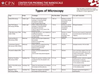
Types of Microscopes • Compound Microscope • Comparison Microscope • Stereoscope Microscope • Polarizing Microscope • Confocal microscopy • Scanning Electron Microscope • Transmission Electron Microscope
Simple Microscope • Light passes through only 1 lens. • Example: magnifying glass
Compound Microscope • Lets light pass through an object and then through two or more lenses.
Compound Light Microscope • A compound light microscope is a microscope with more than one lens and its own light source. In this type of microscope, there are ocular lenses in the binocular eyepieces and objective lenses in a rotating nosepiece closer to the specimen. • Although sometimes found as monocular with one ocular lens, the Compound binocular microscope is more commonly used today. • The first light microscope dates back to 1595, when Zacharias Jansen created a compound microscope that used collapsing tubes and produced magnifications up to 9X. • Microscopes have come a long way since then—today's strongest compound microscopes have magnifying powers of 1,000 to 2,000X.
Compound Light Microscope Uses/Benefits • One of the biggest benefits of owning a light microscope is its simplicity and its convenience. • A compound light microscope is relatively small, therefore it’s easy to use and simple to store, and it comes with its own light source. • Moreover, because of their multiple lenses, compound light microscopes are able to reveal a great amount of detail in samples. • Even an inexpensive one can reveal an incredible view of the world that would be impossible to explore with the naked eye.
Comparison Microscope • The comparison microscope consists of two independent objective lenses joined together by an optical bridge to a common eyepiece lens. • When a viewer looks through the eyepiece lens of the comparison microscope, the objects under investigation are observed side-by-side in a circular field that is equally divided into two parts. • Modern firearms examination began with the introduction of the comparison microscope, with its ability to give the firearms examiner a side-by-side magnified view of bullets.
Applications • This product is an ideal instrument for public security, inspection, court on document retrieval, marks checking and school teaching (used for bullets, tool marks, fingerprints, seals, texts, coins comparison and identification), but also for industries and sectors such as banks, archaeology, electronics, biology, agriculture which need to identify on objects to use. • Comparison microscope XZB-5D successfully solved the technical problems of comparing ridge width and consistency adjustable (this technology is included in the national technology security projects), XZB-5D is an upgraded product improved on XZB-5C model, which not only retains excellent technical performances of XZB-5C, but also increases its image quality and magnification, increases illumination ways and a variety of accessories, shape, structure more beautiful, reasonable, reliable, and makes the instrument fully plays its functions of comparison and identification. Advantages
Stereoscopic Microscope • A stereo, or dissection, microscope is a specialized compound microscope that has two spatially-separated optical paths. • When a user looks through a stereo microscope, the two light paths image the specimen at slightly different angles (parallax) which is interpreted as stereo vision. • Stereo microscopes generally have a lower magnification than high-mag compound microscopes because 1) we manipulate and dissect relatively large objects, and 2) low magnification necessarily has a broader depth of field. • This property is useful for dissections.
Stereoscopic Microscope • Gives a three dimensional view of an object. (Examples: insects and leaves) • Used for dissections
Polarizing Microscopy • Light that is confined to a single plane of vibration is said to be plane- polarized. • The examination of the interaction of plane-polarized light with matter is made possible with the polarizing microscope. • Polarizing microscopy has found wide applications for the study of birefringent materials (materials that split a beam of light in two, each with its own refractive index value). • The determination of these refractive index data provides information that helps to identify minerals present in a soil sample or the identity of a man-made fiber.
Polarizing Microscopes • This can provide information about the shape, color, and size of minerals and it is used to identify hair, human-made fibers and paint. Hair Sample: Polarized Light Hair Sample: Natural Light
Applications of Polarizing Microscope • Geologists use this type of microscope. Geological specimen to be studied is placed on a slide on a rotatable specimen stage. The specimen is then illuminated by a light source under the specimen stage. • They also are being used in medicine. • Polarizing microscopy can be used both with reflected and transmitted light. • Reflected light is useful for the study of opaque materials such as mineral oxides and sulphides, metals and silicon wafers. • Polarizing microscopes are extremely useful for specialized medical and industrial applications, such as identifying crystals or fibers suspended in liquid, identifying minerals in core samples and detecting defects in semiconductors or finding stress points in metal, glass and other materials. • The objectives used in a polarizing microscope are required to be strain free.
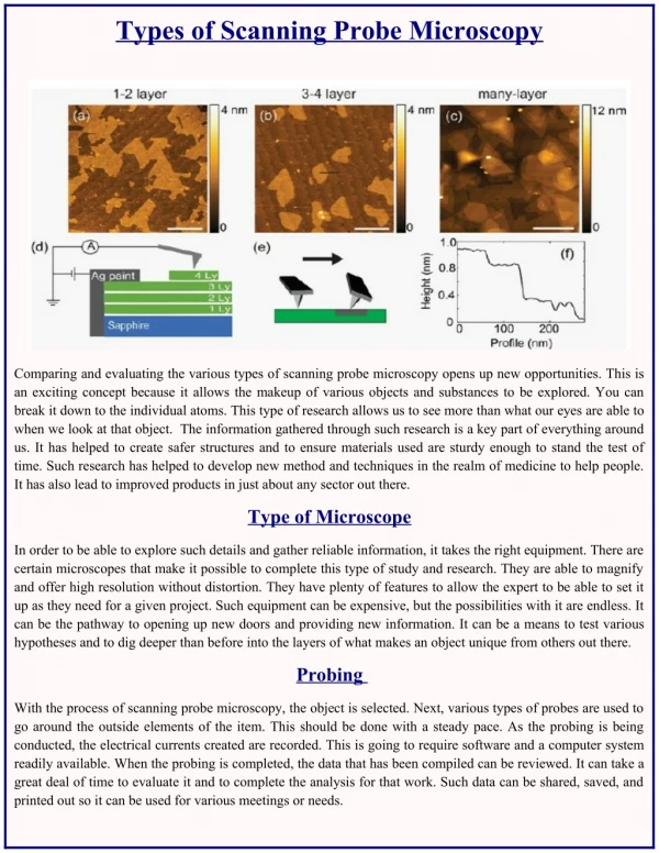
Scanning Electron Microscope(SEM) • A Scanning Electron Microscope (SEM) is a powerful magnification tool that utilizes focused beams of electrons to obtain information. • The high-resolution, three-dimensional images produced by SEMs provide topographical, morphological and compositional information makes them invaluable in a variety of science and industry applications. • The Scanning Electron Microscope developed by professor Dr. Charles Oatlev with the assistance of graduate students in the 1950s, are one of the three types of electron microscopes (EM).
SEM Applications • SEMs have a variety of applications in a number of scientific and industry-related fields, especially where characterizations of solid materials is beneficial. • In addition to topographical, morphological and compositional information, a Scanning Electron Microscope can detect and analyze surface fractures, provide information in microstructures, examine surface contaminations, reveal spatial variations in chemical compositions, provide qualitative chemical analyses and identify crystalline structures. • SEMs can be as essential research tool in fields such as life science, biology, gemology, medical and forensic science, metallurgy. • In addition, SEMs have practical industrial and technological applications such as semiconductor inspection, production line of miniscule products and assembly of microchips for computers.
SEM Advantages and disadvantages • Advantages of a Scanning Electron Microscope include its wide-array of applications, the detailed three-dimensional and topographical imaging and the versatile information garnered from different detectors. • SEMs are also easy to operate with the proper training and advances in computer technology and associated software make operation user-friendly. • The disadvantages of a Scanning Electron Microscope start with the size and cost. • SEMs are expensive, large and must be housed in an area free of any possible electric, magnetic or vibration interference. • Maintenance involves keeping a steady voltage, currents to electromagnetic coils and circulation of cool water. • Special training is required to operate an SEM as well as prepare samples.
Transmission electron microscopy (TEM) • A Transmission Electron Microscope (TEM) utilizes energetic electrons to provide morphologic, compositional and crystallographic information on samples. • At a maximum potential magnification of 1 nanometer, TEMs are the most powerful microscopes. TEMs produce high-resolution, two-dimensional images, allowing for a wide range of educational, science and industry applications. • Ernst Ruska developed the first electron microscope, a TEM, with the assistance of Max Knolls in 1931. After significant improvements to the quality of magnification, Ruska joined the Sieman’s Company in the late 1930s as an electrical engineer, where he assisted in the manufacturing of his TEM.
Transmission electron microscopy (TEM) • Allows the observation of molecules within cells • Allows the magnification of objects in the order of 100, 000’s. • Provides for detailed study of the internal ultrastructure of cells. • a beam of electrons is transmitted through the specimen for a 2D view
Specimen Preparation • analogous to procedures used for light microscopy • for transmission electron microscopy, specimens must be cut very thin • specimens are chemically fixed and stained with electron dense material Other preparation methods • shadowing coating specimen with a thin film of a heavy metal • freeze-etching freeze specimen then fracture along lines of greatest weakness (e.g., membranes)
Transmission electron microscope Chloroplast from a tobacco leaf H1N1 virus
TEM Applications • A Transmission Electron Microscope is ideal for a number of different fields such as life sciences, nanotechnology, medical, biological and material research, forensic analysis, gemology and metallurgy as well as industry and education. • TEMs provide topographical, morphological, compositional and crystalline information. • The images allow researchers to view samples on a molecular level, making it possible to analyze structure and texture. • This information is useful in the study of crystals and metals, but also has industrial applications. • TEMs can be used in semiconductor analysis and production and the manufacturing of computer and silicon chips. • Technology companies use TEMs to identify flaws, fractures and damages to micro-sized objects; this data can help fix problems and/or help to make a more durable, efficient product.
TEM advantages and disadvantages • TEMs offer the most powerful magnification, potentially over one million times or more • TEMs have a wide-range of applications and can be utilized in a variety of different scientific, educational and industrial fields • TEMs provide information on element and compound structure • Images are high-quality and detailed • TEMs are able to yield information of surface features, shape, size and structure • They are easy to operate with proper training • Some cons of electron microscopes include: • TEMs are large and very expensive • Laborious sample preparation • Potential artifacts from sample preparation • Operation and analysis requires special training • Samples are limited to those that are electron transparent, able to tolerate the vacuum chamber and small enough to fit in the chamber • TEMs require special housing and maintenance • Images are black and white
Confocal microscopy • An optical imaging technique for increasing optical resolution and contrast of a micrograph. • Radiations emitted from laser cause sample to fluoresce. • Uses pinhole screen to produce high resolution images. • Eliminates out of focus. • So images have better contrast and are less hazy. • A series of thin slices of the specimen are assembled to generate a 3- dimensinal image. • Is an updated version of fluorescence microscopy.
ADVANTAGES • The specimen is everywhere illuminated axially, rather than at different angles, thereby avoiding optical aberrations. • Entire field of view is illuminated uniformly. • The field of view can be made larger than that of the static objective by controlling the amplitude of the stage movements. Image formed are of better resolution. • Cells can be live or fixed. • Serial optical sections can be collected. • Taking a series of optical slices from different focus levels in the specimen generates a 3D data set. 14
Confocal Laser Scanning Microscope (CLSM) • laser beam used to illuminate spots on specimen • computer compiles images created from each point to generate a 3-dimensional image • used on specimens that are too thick for a light microscope
Applications of Confocal Microscopy • In recent years confocal microscopy has been adapted into numerous fields - such as cell biology, life sciences, semiconductor inspection, materials science, neuroanatomy, and neurophysiology. Key applications areas include: • Stem cell research • Photo bleaching studies • Fluorescence resonance energy transfer (FRET) • Fluorescence recovery after photo-bleaching • Fluorescence in-situ hybridization • Lifetime imaging • Multiphoton microscopy • Total internal reflection fluorescence microscopy (TIRFM) • DNA hybridization • Membrane and ion probes • Bioluminescent proteins • Epitope tagging