Download
1 / 8
80 likes | 190 Vues
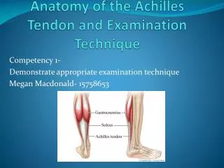
Anatomy of the Achilles Tendon and Examination Technique. Competency 1- Demonstrate appropriate examination technique Megan Macdonald- 15758653. Anatomy.

E N D
Anatomy of the Achilles Tendon and Examination Technique Competency 1- Demonstrate appropriate examination technique Megan Macdonald- 15758653
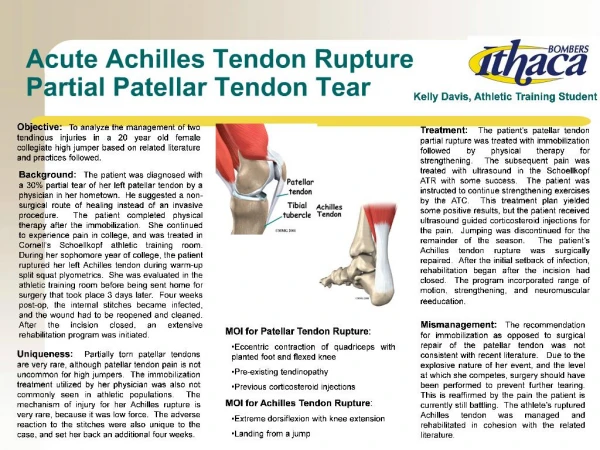
Anatomy • The Achilles tendon is formed by the two tendon heads of the gastrocnemius and soleus muscle. It is considered the strongest tendon within the human body which is located on the posterior aspect of the ankle and is approximately 10-15cm in length (Nunley, 2009). • The tendon is unique in that after the soleous portion the achilles , gastrocnemious and soleal tendon spiral and twist 90 degrees which adds to the strength of the tendon (McNally, 2014). • When in transverse, medial to the Achilles tendon the Plantaris can often be seen. Directly anterior to the achilles is the Kager fat pad. Superior to the distal achillesis the retro- achillesbursa. Inferior to the achillesin close proximity to the calcaneusis the Retro-calcanealbursa (Jacobson, 2013).
Origin • The Achilles Tendon is situated in the middle of the posterior calf. It is formed by the gastrocnemious and soleal tendons combined(Nunley, 2009). www.webmd.com
Insertion • The Achilles tendons inserts on the posterior aspect of the calcanealtuberosity(Nunley, 2009). Normal Achilles tendon in Transverse & Longitudinal http://www.ncbi.nlm.nih.gov/pmc/articles/PMC3495576/figure/bjr-85-1157-g001/
Action • The achilles tendon provides the connection from the muscles to the calceneus allowing plantar flexion of the ankle (Maquirriain, 2011). http://uvahealth.com/services/childrens-hospital/conditions-treatments/203519
Examination Technique • Patient positioning= Patient supine on the table with foot hanging of the end of the bed. Dorsiflexion of the foot aids for better visualisation of the tendon. • Equipment= Use lots of gel to allow appropriate contact. High frequency linear transducer. Typically greater than 10MHz. • Technique= Assessing the tendon in both longitudinal and Transverse beginning at the calcanealinsertion to the myotendinous junction. http://www.essr.org/html/img/pool/ankle.pdf
Ultrasound Appearance • On Ultrasound the normal achillestendon should appear uniform in thickness ,no more than 6mm in the AP dimension (Dong & Fessell, 2009) . • The tendon should consist of a uniform echogenicity with echogenic lines throughout (Bleakney, White, & Maffulli, 2007). Normal Transverse Achilles Tendon
Bibliography • Nunley, J. A. (2009). The Achilles Tendon: Treatment and Rehabilitation. Ney York: Springer. • McNally, E. (2014). Practical Musculoskeletal Ultrasound 2nd edn. New York: Churchill Livingstone. • ESSR. (2013). Musculoskeletal Ultrasound Technical Guidelines VI Ankle. Retrieved 2014, from European Society of MusculoSkeletal Radiology: http://www.essr.org/html/img/pool/ankle.pdf • Dong, Q., & Fessell, D. P. (2009). Achilles Tendon Ultrasound Technique. American Journal of RoentgenologyColume 193 No 3 , 173. • ESSR. (2013). Musculoskeletal Ultrasound Technical Guidelines VI Ankle. Retrieved 2014, from European Society of MusculoSkeletal Radiology: http://www.essr.org/html/img/pool/ankle.pdf • McNally, E. (2014). Practical Musculoskeletal Ultrasound 2nd edn. New York: Churchill Livingstone. • Bleakney, R. R., White, L. M., & Maffulli, N. (2007). Imaging of the Achilles Tendon. Springer. • Jacobson, J. A. (2013). Findamentals of Musculoskeletal Ultrasound. Philadelphia: Elsevier Saunders. • Maquirriain, J. (2011). Achilles Tendon Rupture: Avoiding Tendon Lengthening during Surgical Repair and Rehabilitation. Yale Journal of Biology and Medicine , 289-300.