Bone Anatomy and Skeletal System Overview
Learn about the skeletal system, bone classification, structure, functions, and types of bone tissues in this comprehensive guide. Explore the features of long bones, bone development, and bone marrow roles. Discover the differences between compact and spongy bones.

Bone Anatomy and Skeletal System Overview
E N D
Presentation Transcript
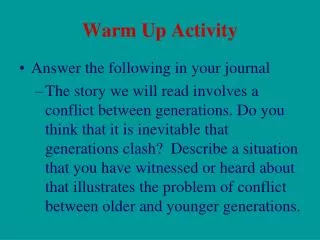
Warm-Up Activity • Fill in the names of the bones in the skeleton diagram.
Warm-Up • What are the 4 types of bones? Give an example of each. • Give 3 ways you can tell a female skeleton from a male skeleton. • What hormones are involved in the skeletal system?
Skeletal System Exercise 8
Axial skeleton: long axis (skull, vertebral column, rib cage) • Appendicular skeleton: limbs and girdles
Axial Skeleton Appendicular Skeleton • Cranium (skull) • Mandible (jaw) • Vertebral column (spine) • Cervical vertebrae • Thoracic vertebrae • Lumbar vertebrae • Sacrum • Coccyx • Sternum (breastbone) • Ribs • Clavicle (collarbone) • Scapula (shoulder blade) • Coxal (pelvic girdle) • Humerus (arm) • Radius, ulna (forearm) • Carpals (wrist) • Metacarpals (hand) • Phalanges (fingers, toes) • Femur (thigh) • Tibia, fibula (leg) • Tarsal, metatarsals (foot) • Calcaneus (heel) • Patella (knee)
Functions of the Bones • Support body and cradle soft organs • Protect vital organs • Movement: muscles move bones • Storage of minerals (calcium, phosphorus) & growth factors • Blood cell formation in red bone marrow • Triglyceride (fat) storage in yellow bone marrow
Classification of Bones • Long bones • Longer than they are wide (eg. femur, metacarpals) • Short bones • Cube-shaped bones (eg. wrist and ankle) • Sesamoid bones (within tendons – eg. patella) • Flat bones • Thin, flat, slightly curved (eg. sternum, skull) • Irregular bones • Complicated shapes (eg. vertebrae, ischium)
Features of a Long Bone • A long bone has two parts: the diaphysis and the epiphysis. • The diaphysis is the hollow tubular shaft between the proximal and distal ends. • The hollow region is called the medullary cavity –filled with yellow bone marrow. • The walls are composed of compact bone.
The wider section at the ends is the epiphysis (plural = epiphyses), composed of spongy bone. Red marrow fills the spaces in the spongy bone. Each epiphysis meets the diaphysis at the epiphyseal plate (growth plate), a layer of hyaline cartilage. • When the bone stops growing in early adulthood (approximately 18–21 years), the cartilage is replaced by osseous tissue and the epiphyseal plate becomes an epiphyseal line.
The medullary cavity is lined with a membrane - endosteum (end- = “inside”; oste- = “bone”), where bone growth, repair, and remodeling occur. • The outer surface of the bone is covered with a fibrous membrane called the periosteum (peri– = “around” or “surrounding”). The periosteum contains blood vessels, nerves, and lymphatic vessels that nourish compact bone. Tendons and ligaments also attach to bones at the periosteum.
The periosteum covers the outer surface except where the epiphyses meet other bones to form joints. In this region, the epiphyses are covered with hyaline cartilage called articularcartilage, that reduces friction and acts as a shock absorber.
Adult = 206 bones • Types of bone tissue: • Compact bone: outer layer – dense & solid • Spongy bone: inner layer - open spaces, red bone marrow • Features: • Very hard (calcium salts) • Light weight • Ability to resist tension and forces (collagen fibers)
Compact Bone The extracellular matrix consists of collagen fibers and a hard ground substance in mostly calcium salts, are deposited. Osteocytes are located in lacunae. Little canals, or canaliculi serve as passageways for nutrients and wastes.
The functional unit of compact bone is called an osteon. • In the center of each osteon is a large central canal, Haversian Canal, which contains blood vessels and nerves; and each osteon is made of concentric ring layers called lamellae. • Canaliculi run perpendicular to the central or Haversian canal and lacunae.
Interstitial lamellae fill in the spaces between osteons in the bone.
Perforating (Volkmann’s) Canals run into compact bone at right angles to the shaft. This allows for communication and transportation of substances to the interior of the bone • Found in the skeleton of all vertebrates.
Ground Compact Bone Lacunae Canaliculi Concentric Lamella Osteon Central Canal Interstitial Lamella
Spongy Bone • Cancellous or trabecular bone is made up of spongy, porous, bone tissue that is filled with red bone marrow. • It is found in the ends(epiphyses) of long bones and in the interior of short, flat and irregular bones (the pelvis, ribs, vertebrae, and skull.)
Spongy bone does not have osteons but instead has trabeculae (little beams) – flat plates with a lattice-like network of thin, bony columns lined with endosteum. • The trabeculae have lamellae, lacunae, osteocytes and canaliculi. • Spongy bone has many spaces filled with red bone marrow.
Trabeculae Spaces with red bone marrow Lacunae with osteocytes
Bone Development • Osteogenesis (ossification): bone tissue formation Stages: • Begins at 8 weeks gestation • Start as cartilage replaced by bone • Post-natal bone growth early adulthood • Epiphyseal plates: (growth plates) regions where long bones lengthen • Appositional growth: bones increase in thickness • Bone modeling and repair – lifelong
“Soft spots” felt on an infant's skull are actually fontanelles • Fibrous connective tissue that connect the incompletely developed flat bones
Hormonal Control • Growth hormones: stimulate longitudinal bone growth • Thyroid hormone: control activity of growth hormone • Testosterone & estrogens(at puberty): • Adolescent growth spurt • Close epiphyseal plates end growth
Bone Cells • Osteoblasts: bone-forming cells • Osteocytes: mature bone cell (doesn’t divide) • Osteoclasts: dissolve/break down bone (bone resorption)
Fractures (Breaks) Classified by: • Position of bone – nondisplaced (normal) or displaced (bone out of alignment) • Completeness of break – complete (broken through) or incomplete • Orientation to long axis of bone – linear (parallel to bone) or transverse (perpendicular to bone) • If bone penetrates skin – open (compound) fracture or closed (simple) fracture
Male Skull Larger and heavier Forehead shorter Face less round Jaw larger Mastoid processes more prominent Male pelvic bones Heavier and thicker Obturator foramen and acetabula are larger and closer together Bone Structure: Gender Differences
Male pelvic cavity Narrower and longer Less roomy and more funnel shaped Male sacrum Narrower Sacral promontory projects forward Sacral curvature is less sharp posteriorly Male coccyx Less movable Bone Structure: Gender Differences
The Skull • 2 bone types: • Cranial – form the top, sides, and back of the skull • Facial – form the face
“Soft spots” felt on an infant's skull are actually fontanelles • Fibrous connective tissue that connect the incompletely developed flat bones
Frontal – anterior Parietal– top and most of the sides Occipital –back Temporal – form the lower sides of the skull Sphenoid and ethmoid bones – floor Ear ossicles are the smallest bones of the body Malleus Incus Stapes The Skull: Cranial Bones
Mandible – forms the lower jaw bone Maxilla – forms the upper jawbone Zygomatic – form the prominence of the cheeks Nasal bones – fuse together to form the bridge of the nose Palatine – form the anterior portion of the hard palate Vomer – a thin bone that divides the nasal cavity The Skull Facial Bones
The Spinal Column • 7 Cervical vertebrae • 12 Thoracic vertebrae • 5 Lumbar vertebrae • Sacrum • Coccyx
Cervical vertebrae Smallest and lightest Located in the neck region C1 = Atlas C2 = Axis Thoracic vertebrae Join the 12 pairs of ribs Lumbar vertebrae Have very sturdy structures Weight-bearing The Spinal Column (cont.)
The Spinal Column (cont.) • Sacrum • Triangular-shaped bone 5 fused vertebrae • Coccyx • Small, triangular bone 3-5 fused vertebrae • Considered unnecessary • Also called the tailbone
ANSWER: Cervical – 7 Apply Your Knowledge Identify the sections of the spinal column and give the number of vertebrae for each. Thoracic – 12 Sacrum – 5 fused Lumbar – 5 Right! Coccyx – 3 to 5 fused