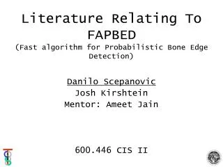
Literature Relating To FAPBED (Fast algorithm for Probabilistic Bone Edge Detection)
430 likes | 597 Vues
Literature Relating To FAPBED (Fast algorithm for Probabilistic Bone Edge Detection). Danilo Scepanovic Josh Kirshtein Mentor: Ameet Jain 600.446 CIS II. Papers Covered. Thoroughly:

Literature Relating To FAPBED (Fast algorithm for Probabilistic Bone Edge Detection)
E N D
Presentation Transcript
Literature Relating To FAPBED(Fast algorithm for Probabilistic Bone Edge Detection) Danilo Scepanovic Josh Kirshtein Mentor: Ameet Jain 600.446 CIS II
Papers Covered Thoroughly: • Jain, Ameet Kumar, Tayor, Russell H., Understanding Bone responses in B-mode Ultrasound Images and Automatic Bone Surface extraction using a Bayesian Probabilistic Framework, SPIE Medical Imaging 2004, Ultrasonic Imaging and Signal Processing. • Zou, Kelly H., et al.Statistical Validation Based on Parametric Receiver Operating Characteristic Analysis of Continuous Classification Data, Acad Radiol 2003; 10:1359-1368.
Papers Covered Summarized: • Muratore, Diane M., et al., Three-Dimensional Image Registration of Phantom Vertebrae for Image-Guided Surgery: A Preliminary Study, Comp Aid Surg 7:342-352 (2002). • Brendel, B., et al., Registration of 3D CT and Ultrasound Detasets of the Spine using Bone Structures, Comp Aid Surg 7:146-155 (2002). • Brendel B. et al., Bone registration with 3D CT and ultrasound data sets, International Congress Series 2192 (2003) 1-7. • Leotta, Daniel F., Martin, Roy W., Three-Dimensional Spatial Compounding of Ultrasound Scans with Weighting by Incidence Angle, Ultrasonic Imaging 22, 1-19 (2000).
Presentation Plan • FAPBED Review • Summary of less-relevant papers • Study of US Paper by Jain • Relevance to FAPBED • Critique • Study of ROC Paper by Zou • Relevance to FAPBED • Critique • FAPBED Future
FAPBED Review Part of a bigger project: Preoperative CT Procedure Plan FAPBED Tracked US Actual Procedure Prob.Bone Surface Prob.Bone Surface CT-US Registration
FAPBED Review Why probabilistic bone surface? The registration-segmentation duality: Our goal is registration, so there is no need to spend time on an accurate segmentation, when we can go ‘directly’ to the goal. Accurate Segmentation Accurate Registration
FAPBED Review • Completed: • Probabilistic edge extraction in US • Pending: • Probabilistic edge extraction in CT • Registration • Concrete FAPBED goal:Fast-running algorithm to give probabilities of being a bone edge for CT data
Presentation Plan • FAPBED Review • Summary of less-relevant papers • Study of US Paper by Jain • Relevance to FAPBED • Critique • Study of ROC Paper by Zou • Relevance to FAPBED • Critique • FAPBED Future
4-Paper Summary 1/4 Three-Dimensional Image Registration of Phantom Vertebrae for Image-Guided Surgery: A Preliminary Study Method:Place 5 posts into each vertebra of phantom spine, segment US images, segment CT images (marching cubes), register using ICP, calculate error using correct transformation
4-Paper Summary 1/4 Results: better registration for higher-resolution CT images (2- and 3-mm slice thickness), better registration for higher-frequency US transducer (7.5MHz), TRE 1.33mm in best case
4-Paper Summary 2/4 Registration of 3D CT and Ultrasound Detasets of the Spine using Bone Structures Method:Threshold CT, estimateedge visible to US given scan path, register CT to unprocessed US by maximizing gray value
4-Paper Summary 2/4 Results: • Registration not sensitive to parameters of surface extraction • The gray value maximization algorithm is robust and can detect misalignments of up to 20º and 40mm:
4-Paper Summary 3/4 Bone registration with 3D CT and ultrasound data sets Method:Threshold CT; extract whole bone surface; estimate surface visible to US given scan path
4-Paper Summary 3/4 Method (cont.):Apply depth gain compensation (DGC) to US; register using maximum gray value, deepest descent Results: Registration takes between 15s and 1 min, converges for initial misalignments of about 30mm and 15º
4-Paper Summary 4/4 Three-Dimensional Spatial Compounding of Ultrasound Scans with Weighting by Incidence Angle Method: compound multiple US images by weighting data based on relation of surface normal and incidence angle
4-Paper Summary4/4 Results: The weighted approach was compared to the maximum and mean compounding approaches, process takes about 100 minutes for 4 scans, slight reduction in edge variability
4-Paper Summary 4/4 Results(cont): biggest advantage for automatic segmentation algorithms
Presentation Plan • FAPBED Review • Summary of less-relevant papers • Study of US Paper by Jain • Relevance to FAPBED • Critique • Study of ROC Paper by Zou • Relevance to FAPBED • Critique • FAPBED Future
The Main Ideas • US is inherently inaccurate and noisy • US contains a lot of partial information: intensity gradients, shadow regions, multiple reflections • Hard segmentation is unnecessary, but a probability of a pixel being an edge can be calculated • The registration should be able to update if more information becomes available (more images) Slide from SPIE presentation by Ameet Kumar Jain
US Inaccuracies • Angle of incidence and bone geometry can skew surface appearance in image Slide from SPIE presentation by Ameet Kumar Jain
US Inaccuracies Exact edge is impossible to deduce,but a region of high likelihood exists Slide from SPIE presentation by Ameet Kumar Jain
Probabilistic Framework • S(i,j) is a random variable. • P(A) is the probability of event ‘A’ • An Image consists of features:{F1,F2,…Fi} ≡ {F}i • A probabilistic feature model P(Fi|S) is usually known Slide from SPIE presentation by Ameet Kumar Jain
Probabilistic Framework • Bayes rule allows us to use what we know in order to deduce what we want • New features can be added to revise probabilities: Slide from SPIE presentation by Ameet Kumar Jain
Probabilistic Framework • Sometimes the model P(Feature| Surface) is too complicated for some feature, but P(Surface| Feature) is extractable. • To couple two surface probabilities • If P1 = P(S|{Fi}) and P2= P(S|{Fj}). P has nice properties Slide from SPIE presentation by Ameet Kumar Jain
Results • Original image(top left),final probability map(top right), probability > 0.5(bottom left),weak segmentation(bottom Right) Slide from SPIE presentation by Ameet Kumar Jain
Results • Feature probability models developed for intensity, shape, multiple reflections • Unoptimized Matlab implementation takes 5s to run on a PC for each image (Pentium4, 2.4GHz and 512MB RAM) • Over 80% of the time is taken to resolve multiple reflections, scanline [re]construction • A 3D US volume construction will remove any remaining artifacts existing in any single image; and sharpen the desired features • True validation would be indicated by the accuracy of the registration algorithm Slide from SPIE presentation by Ameet Kumar Jain
Presentation Plan • FAPBED Review • Summary of less-relevant papers • Study of US Paper by Jain • Relevance to FAPBED • Critique • Study of ROC Paper by Zou • Relevance to FAPBED • Critique • FAPBED Future
Relevance to FAPBED • US and CT images pose similar problems for segmentation • The probabilistic approach to “segmentation” should be less computationally intensive and more information-based than standard CT processing techniques • Bone features can be identified in CT images and the same probability calculations applied to generate the final probability map
Paper Critique • Good • Clearly written, well organized • Not overly mathematical • Detailed enough to convey concepts accurately • Could improve • Some figures were unclear • Reproducing the method might be somewhat difficult given just the information in the paper
Presentation Plan • FAPBED Review • Summary of less-relevant papers • Study of US Paper by Jain • Relevance to FAPBED • Critique • Study of ROC Paper by Zou • Relevance to FAPBED • Critique • FAPBED Future
The Main Ideas • Images can be classified on a discrete or continuous scale • It is difficult to calculate the accuracy of continuous classifications • ROC analysis can be used to evaluate continuous classifiers and provide a value describing their accuracy • AUC of 0.7 or higher indicates fair to excellent accuracy
ROC Analysis • First introduced to evaluate radars in WW2, then expanded to study transistor responses, medical imaging, and data mining Threshold PD Response PFA Text from http://www.dsic.upv.es/~jorallo/Albacete/ROCAnalysis-1.6-English.pdf
ROC Analysis • As the threshold is varied, different values of PD and PFA result, and can be plotted • AUC-area under ROC curve represents classifier accuracy Figure from http://www.dsic.upv.es/~jorallo/Albacete/ROCAnalysis-1.6-English.pdf
Methods • For each disease (MRI of brain tumors, CT of urethral stones, MRI of prostate cancer), determine gold standard • Given classified data and gold standard, find all points that should be diseased and model this distribution (beta)
Methods • Find all points that are not diseased and model the distribution (beta) • Construct ROC curve and calculate AUC • If AUC>0.7, classifier accuracy is fair-excellent
Results • All classifiers were deemed fair-excellent • For example, brain tumor data:
Presentation Plan • FAPBED Review • Summary of less-relevant papers • Study of US Paper by Jain • Relevance to FAPBED • Critique • Study of ROC Paper by Zou • Relevance to FAPBED • Critique • FAPBED Future
Relevance to FAPBED • We have raw and hand-segmented CT images (gold standard) • After running FAPBED on these images, ROC analysis can be conducted in a similar way • The AUC can be used as an evaluation of accuracy • If hard-segmenting is desirable, the optimal threshold can be determined • Relative AUC data can be used to speed up algorithm if additional features increase processing time but not accuracy
Paper Critique • Good • Very detailed, easily replicated • Simple but useful idea • Bad • Overly mathematical, gets in the way of concepts • Many typographical errors • Didn’t try hard enough?
Presentation Plan • FAPBED Review • Summary of less-relevant papers • Study of US Paper by Jain • Relevance to FAPBED • Critique • Study of ROC Paper by Zou • Relevance to FAPBED • Critique • FAPBED Future
FAPBED Future • Incorporate probabilistic methods discussed in Jain paper • Use ROC to evaluate performance of various methods as shown in Zou paper • Possible to produce optimizedprogram, where speed and accuracy are maximized • Try alternate registration methods, like gray-level maximization
Thanks I would like to thank Josh Kirshtein for doing a thorough literature search and finding these articles, and Ameet Jain for explaining his paper and the main ideas of the other papers, and providing the presentation slides and pictures for his paper Questions?