Download
1 / 55
550 likes | 577 Vues
Learn about acute rheumatic fever (ARF) and rheumatic heart disease (RHD), including epidemiology, diagnosis, Jones criteria, valvular heart disease, and prevention strategies. Explore the impact, burden, and clinical manifestations of these conditions. This lecture provides valuable insights for healthcare professionals.

E N D
Rheumatic Fever And RHD Dr. Abdulelah Mobeirek (FRCPC) Consultant Cardiologist KFCC
Lecture Outline • What is ARF And RHD? • Diagnosis • Jones Criteria • Differential Diagnosis • Investigations, Management • Rheumatic Valvular Heart Disease • Prevention
Rhuematic Fever • Follows group A beta hemolytic streptococcal throat infection • It represents a delayed immune response to infection with manifestations appearing after a period of 2-4 weeks • Age 5-15 yrs • A multisystem disease • RHD is a long term complication og ARF • Major effect on health is due to damage to heart valves
Pathologic Lesions • Ashcoff nodules: • Fibrinoiddegeneration of connective tissue, inflammatory cells
Global Burden of RHD • Total cases with RHD:20 Millions • CHF: 3 Million • Valve surgery required in 1 Million • Annual incidence of RF: 0.5 Million, nearly half develop carditis • Estimated deaths from RHD: 230,000/YR • Imposes a substantial burden on health care systems with limited budgets
Epidemiologic Background • Globally RHD is the commonest CVD in young people 25 yrs old • The overall incidence of ARF from 5-51per 100000 population with a mean of 19 per 100000 population • In children 5-14 yrs old 0.8-5.7 per 1000 children with a median of 1.3 per 1000
Epidemiologic Background • The incidence of RF and the prevalence of RHD has declined substantially in Europe, North America and other developed nations • this decline has ben attributed to improved hygiene, reduced household crowding, and improved medical care
Epidemiologic Background • The major burden is currently found in low and middle income countries, and in selected indigenous populations of certain developed countries. • A disease of poverty and low socioeconomic status • In underdeveloped countries RHD is the leading cause of CV death during the first five decades of life
Diagnosis of ARF • No single test to diagnose ARF • The symptoms and signs are shared by many inflammatory and infectious diseases • Accurate diagnosis is important • Overdiagnosis will result in individuals receiving treatment unnecessarily • Underdiagnosis may lead to further episodes of ARF causing damage, and the need for valve surgery, and or premature death
Diagnosis of ARF • Diagnosis is primarily clinical and is based on a constellation of signs and symptoms, which were initially established as the Jones criteria • In 1944 Dr. TD Jones published a set of guidelines for diagnosis of ARF “Jones Criteria” • Subsequently Modified in 1965, 1984 and 1992by AHA • Revised recently -2015 by AHA
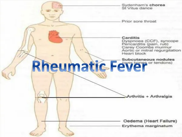
Carditis • Occurs in 50-70% of cases • Only manifestation of ARF that leaves permanent damage • May be subclinical • Murmurs of MR or AR may occur in acute stage while mitral stenosis occurs in late stages • Cardiomegaly and CHF may occur
Arthritis • Common: present in 35-66% • Earliest manifestation of ARF • Large joints: The knees and ankles, shoulders, elbows • “Migrating”, “Fleeting” polyarthritis • Duration short < 1 week • Rapid improvement with salicylates • Does not progress to chronic disease
Sydenham Chorea • Also known as Saint Vitus’dance • Occur in 10-30%, extrapyramidal manifestation, female predominnce • Abrupt Purposeless involuantry movements of muscles of face, neck, trunk, and limbs. • Delayed manifestation of ARF -months • Clinically manifest as-clumsiness, deterioration of handwriting,emotional lability or grimacing of face
Subcutaneous Nodules • Occur in 10% • Usually 0.5 – 2 cm long • Firm non-tender • Occur over extensor surfaces of joints, on bony prominences, tendons, spine • Short lived: last for few days • Associated with severe carditis
Erythema Marginatum • Present in <6% • Less common, but highly specific manifestation of ARF • Reddish border, pale center, round or irregular serpiginous borders, non-pruritic, transient rash • Occurs on trunk, abdomen or proximal limbs • Associated with carditis
2015 Revision of Jones Criteria • In accordance with the degree of prevalence of ARF/RHD in the population: • low risk populations have been defined as those with ARF incidence < 2:100000 school-age children or all age prevalence of RHD of < 1:1000 population per year • Children not from low risk population have been considered to be at moderate or high risk
2015 Revision of Jones Criteria 2. Advocated the use of Echocardiography in all cases of confirmed or suspected ARF or RHD, to diagnose valvulitis( subclinical carditis) and has been included as a major criterion to diagnose carditis 3. Aseptic monoarthritis has been included as a major criteria in moderate or high risk population
2015 Revision of Jones Criteria 4. Polyarthralgiahas been recognized as a major manifestation for moderate or high risk population 5. Fever >38.5 c, ESR >60 and or CRP > 3mg/dl for low risk population, and fever >38 and ESR >30 and or CRP > 3mg/dl for moderate or high risk population
2015 Revised Jones Criteria A firm diagnosis requires • 2 Major manifestations or 1 Major and 2 Minor manifestations and 2 ) Evidence of a recent streptococcal infection.
2015 Revised Jones Criteria Evidence of Preceding GAS Infection: • Increased or rising ASO titer or Anti-Dnase B titer • A positive throat culture
Treatment of ARF • Bed rest • Salicylates : Aspirin • 75-100 mg /kg/day given as 4 divided doses for 6 -8 weeks • Attain a blood level 20-30 mg/dl • Penicillin: Procaine Penicillin 4 million units/day x10 days • Prednisolone:2mg/kg/day taper over 6 weeks, Given when there is severe carditis • Heart Failure Treatment: diuretics, ACEI
Rheumatic Heart Disease • Most commonly in Mitral-70% • Frequently in Aortic-40% • Less frequently Tricuspid-10% • Rarely pulmonary valve-2% • Mitral Stenosis is more common in females(3:1), while males have higher incidence of Aortic Regurgitation
Mitral Stenosis • The normal MVA= 4-6 cm2 • In severe ms<1.5 cm2 • High LAP • The rise in LAP causes a similar rise in pulmonary capillaries, veins and artery
Clinical Features • Dyspnea • Fatigue • Palpitation • Hemoptysis (10%) • Hoarseness ( Ortner’s syndrome) • Dysphagia • Storke or peripheral embolization
Clinical Features • Cyanosis (Mitral facies,malar flush) • Tapping apex ( S1) • Parasternal heave • Diastolic thrill • Accentuated S1 , accentuated S2 • Opening snap • Mid-diastolic rumble
Investigations • CXR • Straightening of the left heart border • Double density • KerleyB lines , CA in MV • ECG: LAE, P Mitrale,RV dominance • Echodoppler
Management • B-Blockers ,CCB • Digoxin ( AF ) • Warfarin • Balloon Valvuloplasty • Mitral valve replacement
Mitral Regurgitation • Asymptomatic • Dyspnea , orthopnea, PND • Displaced PMI, Thrill • Soft S1, • Pansystolic murmur • Treatment is surgical
Aortic Regurgitation • Water-hammer / collapsing pulse • Wide pulse pressure • Corrigan’s sign • De Musset sign • Muller sign • Quincke’s pulse • Hill’s sign
Symptoms • Angina • Syncope • Dyspnea
Signs • Arterial Pulse wave form : Plateau • Small (Parvus) • Slow rise (Tardus) • Sustained not displaced PMI • Systolic thrill • S4
Signs • Late peaking of murmur • Single S2 : Soft or absent A2 • Paradoxical splitting of S2
Aortic Valve Disease Treatment: • Aortic valve Replacement • Transcathter Aortic Valve Replacement