Download
1 / 17
170 likes | 419 Vues
اعداد الدكتور زيد علي مجيد جراح اختصاص Lec . 2. GASTRITIS The understanding of gastritis has increased markedly following elucidation of the role of H. pylori in chronic gastritis. Type A gastritis

E N D
اعداد الدكتور زيد علي مجيد جراح اختصاص Lec. 2
GASTRITIS The understanding of gastritis has increased markedly following elucidation of the role of H. pylori in chronic gastritis. Type A gastritis This is an autoimmune condition in which there are circulating antibodies to the parietal cell. This results in the atrophy of the parietal cell mass, resulting in hypochlorhydria and ultimately achlorhydria. As intrinsic factor is also produced by the parietal cell there is malabsorption of vitamin B12, which, if untreated, may result in pernicious anaemia. In type A gastritis, the antrum is not affected and the hypochlorhydria leads to the production of high levels of gastrin from the antral G cells. This results in hypergastrinaemia. This, in turn, results in hypertrophy of the ECL cells in the body of the stomach, which are not affected by the autoimmune damage. Over time it is apparent that microadenomas develop in the ECL cells of the stomach, sometimes becoming identifiable tumour nodules. Very rarely, these tumours can become malignant. Patients with type A gastritis are predisposed to the development of gastric cancer, andscreeningsuch patients endoscopically may be appropriate. Type B gastritis There are abundant epidemiological data to support the association of this type of gastritis with H. pylori infection. Most commonly, type B gastritis affects the antrum, and it is these patients who areprone to peptic ulcer disease. Helicobacter-associated pangastritisis also a very common manifestation of infection, but gastritis affecting the corpus alone does not seem to be associated. However, there are some data to suggest that Helicobacter may be involved in the initiation of the process. Patients with pangastritisseem to be most prone to the development of gastric cancer. Intestinal metaplasia is associated with chronic pangastritiswith atrophy. Although intestinal metaplasia per se is common, intestinal metaplasia associated with dysplasia has significant malignant potential and, if this condition is identified, endoscopic screening may be appropriate.
Reflux gastritis This is caused by enterogastric reflux and is particularly common after gastric surgery. Its histological features are distinct from those of other types of gastritis. Although commonly seen after gastric surgery, it is occasionally found in patients with no previous surgical intervention or who have had a cholecystectomy. Erosive gastritis This is caused by agents that disturb the gastric mucosal barrier; NSAIDs and alcohol are common causes. The NSAID-induced gastric lesion is associated with inhibition of the cyclo-oxygenasetype 1 (COX-1) enzyme, hence reducing the production of cytoprotectiveprostaglandins in the stomach. Many of the beneficial anti-inflammatory activities of NSAIDs are mediated by COX-2, and the use of specific COX-2 inhibitors reduces the incidence of these side-effects. However, taken in the long term, COX-2 inhibitors appear to be associated with cardiovascular complications, in common with many NSAIDs. Stress gastritis This is a common sequel of serious illness or injury and is characterisedby a reduction in the blood supply to superficial mucosa of thestomach. Although common, it is not usually recognisedunlessstress ulceration and bleeding supervene, in which case Treatment can be extremely difficult. The condition also sometimes follows cardiopulmonary bypass. Prevention of the stress bleedingfrom the stomach is much easier than treating it, hence theroutine use of H2-receptor antagonists, with or without barrier agents such as sucralfate, in patients who are in intensive care. These measures have been shown to reduce the incidence of bleeding from stress ulceration. Ménétrier’s disease This is an unusual condition characterised by gross hypertrophy of the gastric mucosal folds, mucus production and hypochlorhydria. The condition is pre-malignant and may present with hypoproteinaemiaand anaemia. There is no treatment other thana gastrectomy.
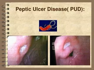
Lymphocytic gastritis This type of gastritis is seen rarely. It is characterised by the infiltration of the gastric mucosa by T cells and is probably associated with H. pyloriinfection. The pattern of inflammation resembles that seen in coeliac disease or lymphocytic colitis. PEPTIC ULCER DISEASE Peptic ulcers are focal defects in the gastric or duodenal mucosa that extend into the submucosa or deeper. They may be acute or chronic and, ultimately, are caused by an imbalance between mucosal defenses and acid/peptic injury . Peptic ulcer remains a common outpatient diagnosis, but the number of physician visits, hospital admissions, and elective operations for PUD has decreased steadily and dramatically over the past three decades. Interestingly, the start of these trends all predated the use of H2 receptor blockers, or proton pump inhibitors, fiberoptic endoscopy, and highly selective vagotomy. However, the incidence of emergency surgery and the death rate associated with peptic ulcers has not decreased nearly so dramatically. PUD is one of the most common GI disorders in the United States with a prevalence of about 2%, and a lifetime cumulative prevalence of about 10%, peaking around age 70 years. The costs of PUD, including lost work time and productivity, are estimated to be above $8 billion per year in the United States. In 1998, approximately 1.5% of all Medicare hospital costs were spent treating PUD. Common sites for peptic ulcers are the first part of the duodenum and the lesser curve of the stomach, but they also occur on the stoma following gastric surgery, the oesophagus and even in a Meckel’s diverticulum, which contains ectopic gastric epithelium. In general, the ulcer occurs at a junction between different types of epithelium, the ulcer occurring in the epithelium least resistant to acid damage. Pathology Most duodenal ulcers occur in the first part of the duodenum . A chronic ulcer penetrates the mucosa and into the muscle coat, leading to fibrosis. The fibrosis causes deformities such as pyloric stenosis. When an ulcer heals, a scar can be observed in the mucosa. Sometimes there may be more than one duodenal ulcer.
The situation in which there is both a posterior and an anterior duodenal ulcer is referred to as ‘kissing ulcers’. Anteriorly placed ulcers tend to perforate and, in contrast, posterior duodenal ulcers tend to bleed, sometimes by eroding a large vessel such as the gastroduodenalartery. Occasionally, the ulceration may be so extensive that the entire duodenal cap is ulcerated and devoid of mucosa. With respect to the giant duodenal ulcer, malignancy in this region is so uncommon that, in normal circumstances, surgeons can be confident that they are dealing with benign disease, even though from external palpation it may not appear so. In the stomach the situation is different. Gastric ulceration Incidence As with duodenal ulceration, H. pylori and NSAIDs are the important aetiological factors in gastric ulceration. Gastric ulceration is also associated with smoking; other factors are of lesser importance. Acid Secretion and Peptic Ulcer In patients with gastric ulcer, acid secretion is variable. Currently, five types of gastric ulcer are described, although the original Johnson classification contained three types . The most common, Johnson type I gastric ulcer, is typically located near the angularisincisura on the lesser curvature, close to the border between antral and corpus mucosa. Patients with type I gastric ulcer usually have normal or decreased acid secretion. Type II gastric ulcer is associated with active or quiescent duodenal ulcer disease, and type III gastric ulcer is prepyloriculcer disease. Both type II and type III gastric ulcers are associated with normal or increased gastric acid secretion. Type IV gastric ulcers occur near the GE junction, and acid secretion is normal or below normal. Type V gastric ulcers are medication induced and may occur anywhere in the stomach. Patients with gastric ulcers may have weak mucosal defenses that permit an abnormal amount of injurious acid back-diffusion into the mucosa. Duodenogastric reflux may play a role in weakening the gastric mucosal defenses, and a variety of components in duodenal juice, including bile, lysolecithin, and pancreatic juice, have been shown to cause injury and inflammation in the gastric mucosa. NSAIDs and aspirin have similar effects. Although chronic gastric ulcer usually is associated with surrounding gastritis, it is unproven that the latter leads to the former.
Patients taking NSAIDs or aspirin need concomitant acid suppressing medication if any of the following risk factors is present : • Age over 60 • History of acid/peptic disease • Concurrent steroid intake • Concurrent anticoagulant intake • High-dose NSAID or acetylsalicylic acid Clinical features of peptic ulcers Pain The pain is epigastric, often described as gnawing, and may radiate to the back. Eating may sometimes relieve the discomfort. The pain is normally intermittent rather than intractable. Periodicity One of the classic features of untreated peptic ulceration is periodicity. Symptoms may disappear for weeks or months to return again. This periodicity may be related to the spontaneous healing of the ulcer. Vomiting Although this occurs, it is not a notable feature unless stenosis has occurred. Alteration in weight Weight loss or, sometimes, weight gain may occur. Patients with gastric ulceration are often underweight but this may precede the occurrence of the ulcer. Bleeding All peptic ulcers may bleed. The bleeding may be chronic and presentation with anaemia is not uncommon. Acute presentation with haematemesis and melaena is discussed later.
Investigation of the patient with suspected peptic ulcer Gastroduodenoscopy This is the investigation of choice in the management of suspected peptic ulceration and, in the hands of a well-trained operator, is highly accurate. In the stomach, any abnormal lesion should be multiply biopsied and, in the case of a suspected benign gastric ulcer, numerous biopsies must be taken to exclude, as far as possible, the presence of a malignancy. Commonly, biopsies of the antrum will be taken to see whether there is histological evidence of gastritis and h. pylori infection. 24 hours acid secretion,, urea breath test for H.pylori infection,, barium meal .. And other investigations. Treatment of peptic ulceration Medical treatment It is reasonable that a doctor managing a patient with an uncomplicated peptic ulcer should suggest modifications to the patient’s lifestyle, particularly the cessation of cigarette smoking. This advice is rarely followed and pharmacological measures form the mainstay of treatment. H2-receptor antagonists (Black) revolutionised the management of peptic ulceration; most duodenal ulcers and gastric ulcers can be healed by a few weeks of treatment with these drugs provided that they are taken and absorbed. There remain, however, a group of patients who are relatively refractory to conventional doses of H2-receptor antagonists. This is largely now irrelevant as proton pump inhibitors can effectively render a patient achlorhydric and all benign ulcers will heal using these drugs, the majority within 2 weeks. Eradication therapy Eradication therapy is now routinely given to patients with peptic ulceration. Evidence suggests that if a patient has a peptic ulcer and H. pylori is the principal aetiologicalfactor (essentially the patient not taking NSAIDs), complete eradication of the organism will cure the disease and reinfection as an adult is uncommon.
Surgical treatment of uncomplicated peptic ulceration Operations for duodenal ulceration Billroth I & II gastrectomy
Gastrojejunostomy Because of the potential for mortality after gastrectomy, the use of gastrojejunostomyalone in the treatment of duodenal ulceration was developed . Reflux of alkali from the small bowel into the stomach reduced duodenal acid exposure and was often successful in healing the ulcer. However, because the jejunal loop was exposed directly to gastric acid, stomal ulceration was extremely common, hence the procedure in isolation was ineffective.
Truncalvagotomy and drainage Highly selective vagotomy Truncalvagotomy and antrectomy
Sequelae of peptic ulcer surgery Recurrent ulceration Although mentioned first, this is by far the easiest problem to treat. Just as all peptic ulcers will heal with potent anti-secretory agents, so will ulcers that are recurrent after ulcer surgery. As with other peptic ulcers, recurrent ulcers may present with complications, particularly bleeding and perforation. In this respect, the complication of gastrojejunal colic fistula requires a particular mention. In this rare condition, the anastomotic ulcer penetrates into the transverse colon. Patients suffer from diarrhoea that is severe and follows every meal. They have foul breath and may vomit formed faeces. Severe weight loss and dehydration are rapid in onset and, for this reason, the condition may be mistaken for malignancy. The major factor producing the nutritional disturbance is the severe contamination of the jejunum with colonic bacteria. A number of imaging techniques can be used to detect the fistula, most commonly CT with oral contrast or, indeed, a barium enema. Endoscopy may not convincingly demonstrate the fistula and, in about one-half of such cases, the barium meal will not reveal the problem. The treatment of gastrocolic fistula consists of first correcting the dehydration and malnutrition and then performing revisional surgery. Small stomach syndrome Early satiety follows most ulcer operations to some degree, including highly selective vagotomy, in which, although there is no anatomical disturbance of the stomach, there is a loss of receptive relaxation. Fortunately, this problem does tend to get better with time and revisional surgery is not necessary. Bile vomiting Bile vomiting can occur after any form of vagotomy with drainage or gastrectomy. Commonly, the patient presents with vomiting of a mixture of food and bile or sometimes bile alone after a meal. Often, eating will precipitate abdominal pain and reflux symptoms are common. Bile-chelating agents can be tried but are usually ineffective. In intractable cases, revisional surgery may be indicated. The nature of the revision depends very much on the original operation.
Following gastrectomy, Roux-en-Y diversion is probably the best treatment. In patients with a gastroenterostomy, the drainage may be taken down and, in most circumstances, a small pyloroplasty can be performed. In patients with a pyloroplasty, reconstruction of the pylorus has been attempted but, in general terms, the results have been rather poor. Antrectomy and Roux-en-Y reconstruction may be the better option. Early and late dumping Although considered together because the symptoms are similar, early and late dumping have different aetiologies A common feature, however, is early rapid gastric emptying. Many patients have both early and late dumping. Early dumping Early dumping consists of abdominal and vasomotor symptoms, which are found in about 5–10% of patients following gastrectomy or vagotomy and drainage. It also affects a small percentage of patients following highly selective vagotomy because of the loss of receptive relaxation of the stomach. The small bowel is filled With foodstuffs from the stomach, which have a high osmotic load, and this leads to the sequestration of fluid from the circulation into the gastrointestinal tract. This can be observed by the rise in the packed cell volume while the symptoms are present. All of the symptoms shown in can be related to this effect on the gut and the circulation. Treatment The principal treatment is dietary manipulation. Small, dry meals are best, and avoiding fluids with a high carbohydrate content also helps. Fortunately, following operation, the syndrome tends to improve with time. For some reason, however, There is a group of patients who suffer intractable dumping regardless of any of these measures. The somatostatin analogue octreotide, given before meals, has been shown to be useful in some individuals and the long-acting preparation may also be useful. However, this treatment can lead to the development of gallstones and it does not help the diarrhoea from which many patients with dumping also suffer. Revisional surgery may be occasionally required. In patients with a gastroenterostomy, the drainage may be taken down or, in the case of a pyloroplasty, repaired. Alternatively, antrectomy with Roux-en-Y reconstruction is often effective, although the procedure is of greater magnitude; following gastrectomy, it is the revisionalprocedure of choice.
Late dumping This is reactive hypoglycaemia. The carbohydrate load in the small bowel causes a rise in the plasma glucose level, which, in turn, causes insulin levels to rise, causing a secondary hypoglycaemia. This can be easily demonstrated by serial measurements of blood glucose in a patient following a test meal. The treatment is essentially the same as for early dumping. Octreotideis very effective in dealing with this problem. Post-vagotomydiarrhoea This can be the most devastating symptom to afflict patients having peptic ulcer surgery. Most patients will suffer looseness of bowel action to some degree (with the exception of highly selective vagotomy) but, in about 5%, it may be intractable. Despite much investigation, the precise aetiology of the problem is uncertain. It is partly related to rapid gastric emptying. In all probability, the denervation of the upper gastrointestinal tract as a result of the vagotomy is also important. Exaggerated gastrointestinal peptide responses may also aggravate the condition. The diarrhoea may take several forms. It may be severe and explosive, with the patient experiencing a considerable degree of urgency. The patient sometimes describes the diarrhoea as feeling like passing boiling water. At the other extreme, some patients only have minor episodes of diarrhoea, which are not as directly related to food. The condition is difficult to treat. The patient should be managed as for early dumping and anti-diarrhoealpreparations may be of some value. Octreotide is not effective in treating this condition and the results of revisional surgery are too unpredictable to make this an attractive option. Malignant transformationPepticulcers Many large studies now confirm that operations such as gastrectomy or vagotomy and drainage are independent risk factors for the development of gastric cancer. The increased risk appears to be approximately four times that of the control population. It is not difficult to understand the increased incidence of gastric cancer, as bile reflux gastritis, intestinal metaplasia and gastric cancer are linked. The lag phase between operation and the development of malignancy is at least 10 years. Highly selective vagotomydoes not seem to be associated with an increased incidence.
Nutritional consequences Nutritional disorders are more common after gastrectomy than after vagotomy and drainage. Weight loss is common after gastrectomy and the patient may never return to their original weight. Eating small meals often may help. Anaemiamay result from either iron or vitamin B12 deficiency. Iron-deficiency anaemiaoccurs after both gastrectomy and vagotomyand drainage and is multifactorial in origin. Reduced iron absorption is probably the most important factor, although the loss of blood from the gastric mucosa may also be important. Vitamin B12 deficiency is prone to occur after total gastrectomy. However, because of the very large vitamin B12 stores, this may be very late in occurring. Vitamin B12 supplementation after total gastrectomy is essential. Rarely, vitamin B12 deficiency may occur after lesser forms of gastrectomy. The cause is a combination of reduced intrinsic factor production and, in destruction of vitamin B12. Bone disease is seen principally after gastrectomy and mainly in women. The condition is essentially indistinguishable from the osteoporosis commonly seen in post-menopausal women; it is only the frequency and magnitude of the disorder that distinguish it. Treatment is through dietary supplementation with calcium and vitamin D, and with exercise. Gallstones The development of gallstones is strongly associated with truncalvagotomy, in which the biliary tree as well as the stomach is denervated, leading to stasis and, hence, stone formation. Patients developing symptomatic gallstones will require cholecystectomy. However, this may induce or worsen other post-peptic Ulcer surgery syndromes such as bilious vomiting and postvagotomydiarrhoea.