Viruses in water
820 likes | 987 Vues
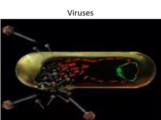
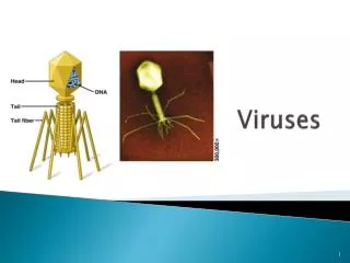
Viruses in water. John Scott Meschke Email: jmeschke@u.washington.edu. Viruses. Small in size (0.02-0.3 micrometers diameter Spherical (icosahedral) or rod-shaped (helical) No biological activity outside of host cells/or host organisms

Viruses in water
E N D
Presentation Transcript
Viruses in water John Scott Meschke Email: jmeschke@u.washington.edu
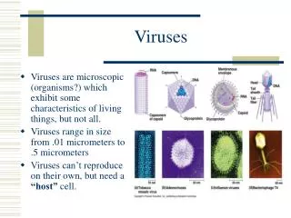
Viruses • Small in size (0.02-0.3 micrometers diameter • Spherical (icosahedral) or rod-shaped (helical) • No biological activity outside of host cells/or host organisms • Obligate intracellular parasites; recruit host cell to make new viruses, often destroying the cell • Non-enveloped viruses tend to be the most persistent in the environment (particularly in aqueous systems) • Protein coat confers stability • Enteric viruses are most relevant for waterborne exposures • Although viruses, spread by other routes, may be present in water samples typical enteric virus E. coli 0.45m Filter
Virion Composition • Nucleic acid: • DNA or RNA • single or double-stranded • 1 or several segments • Capsid (protein coat): • multiple copies of 1 or more proteins in an array • Envelope: • lipid bilayer membrane + glycoproteins) • typically acquired from host cell membranes
Viral Gastroenteritis • It is thought that viruses are responsible for up to 3/4 of all infective diarrhoeas. • Viral gastroenteritis is the second most common viral illness after upper respiratory tract infection. • In developing countries, viral gastroenteritis is a major killer of infants who are undernourished. Rotaviruses are responsible for half a million deaths a year. • Many different types of viruses are found in the gut but only some are associated with gastroenteritis
Viruses found in the gut (1) A. Associated with gastroenteritis • Rotaviruses • Adenoviruses 40 41 • Caliciviruses • Norwalk like viruses or SRSV (Small Round Structured Viruses) • Astroviruses • SRV (Small Round Viruses) • Coronaviruses • Toroviruses
Viruses found in the gut (2) B. Found in the gut, not normally associated with gastroenteritis • Polio • Coxsackie A • Coxsackie B • Echo • Enteroviruses 68-71 • Hepatitis A • Hepatitis E • Adenoviruses 1-39 • Reoviruses C. Found in the gut as opportunistic infection • CMV • HSV • VZV • HIV
Genus: Enterovirus • Icosahedral shape, ~27-30 nm diameter • single-stranded +sense RNA • about 7,500 nucleotides • icosahedral protein coat (capsid) • 4 capsid proteins: VP1, VP2, VP3, VP4 (all cleaved from VP0) • 10 viral species • Human enterovirus A • Human enterovirus B • Human enterovirus C (Polioviruses) • Human enterovirus D • (Human rhinovirus A) • (Human rhinovirus B) • Bovine enterovirus • Porcine enterovirus A • Porcine enterovirus B • Simian enterovirus A
Genus: Rotavirus • ~spherical; icosahedral • ~75-80 nm diameter • double-layered capsid • nucleic acid: • double-stranded RNA • 11 segments rota) • electropherotypes • 5 Species • Human rotavirus A • Human rotavirus B • Human rotavirus C • Human rotavirus D • Human rotavirus E • Other viruses in Reoviridae
Rotaviruses • account for 50-80% of all cases of viral gastroenteritis • usually endemic, but responsible for occasional outbreaks • causes disease in all age groups but most severe symptoms in neonates and young children. Asymptomatic infections common in adults and older children. Symptomatic infections again common in people over 60 • up to 30% mortality rate in malnourished children, responsible for up to half a million deaths per year
Rotaviruses • 80% of the population have antibody against rotavirus by the age of 3 • more frequent during the winter • faecal-oral spread. ? respiratory droplets • 24-48 hr incubation period followed by an abrupt onset of vomiting and diarrhoea, a low grade fever may be present. • Live attenuated vaccines now available for use in children
Genus: Mastadenovirus: • icosahedral • ~80 nm diameter • double-stranded, linear DNA • protein coat contains at least 10 proteins • 6 species (Human adenovirus A-F) • >50 human adenoviruses • mostly respiratory • but may be fecally shed • types 40 and 41 are enteric • Often the most prevalent viruses in treated sewage • resistance to treatment? • Distinct animal adenoviruses
Genus: Norovirus • Icosahedral • “structured”; cup-like surface morphology • 27-35 nm diameter • ss(+) RNA, ~7.7 KB • 1 major capsid polypeptide, ~60 kD • minor protein, ~30 kD • 5 major Norovirus groups, • No culture (except in humans) • Distinct animal Noroviruses
GI.1_Norwalk GI.2_Southampton GI.4_Chiba GI GI.5_Musgrove GI.6_Hesse GI.3_Desert Shield GI.7_Winchester GIII GI.8_Boxer GIII.1_Jena GIII.2_CH126 GII.1_Hawaii GII.12_Wortley GII.16_Tiffin GII.2_Melksham GII.5_Hillingdon GII.10_Erfurt GII.13_Fayetteville GII.17_CS-E1 GII.3_Toronto GII.6_Seacroft GII.7_Leeds GII.8_Amsterdam GII.9_Virginia Beach GII.14_M7 GII GII.11_SW918 GII.4_Bristol GIV GII.15_J23 GV GIV.1_Alphatron GV.1_Murine .10 Genotypes: ex. 29 Clusters of Noroviruses Clusters differ by ≥ 20% amino acid pairwise distance Genogroups differ by 44-55% amino acid pairwise distance
Genus: Hepatovirus • 1 species, Hepatitis A virus • Single serotype worldwide • Acute disease and asymptomatic infection • No chronic infection • Protective antibodies develop in response to infection - confers lifelong immunity
Concentration of Hepatitis A Virus in Various Body Fluids Feces Serum Body Fluids Saliva Urine 102 104 100 106 108 1010 Infectious Doses per mL Source: Viral Hepatitis and Liver Disease 1984;9-22 J Infect Dis 1989;160:887-890
Genus: Hepevirus • 1 species, Hepatitis E virus • Icosahedral • Incubation period: Average 40 days (Range 15-60 days) • Case-fatality rate: Overall, 1%-3%; Pregnant women, 15%-25% • Illness severity: Increased with age • Chronic sequelae: None identified • Most outbreaks associated with fecally contaminated drinking water • U.S. cases usually have history of travel to HEV-endemic areas
Geographic Distribution of Hepatitis E Outbreaks or Confirmed Infection in >25% of Sporadic Non-ABC Hepatitis
The Challenge of Environmental Sampling for Viruses • Variation in virus type and distribution • Low viral numbers: need to concentrate them • Non-random distribution and physical state of viruses of interest: aggregated, particle-associated, embedded, etc. • Volume considerations • Environmental factors may inhibit or interfere with downstream detection • Separate them from interfering and excess other material
Detection of Viruses in The Environment • Three main steps: • (1) recovery and concentration, • (2) purification and separation, and • (3) assay and characterization.
Pathogens in Raw Sewage • Viruses (105-106) • Salmonella (5,000-80,000) • Giardia (9,000-200,000) • Cryptosporidium (1-4000) • In Biosolids: • Viruses ~102-104 (primary) ~102 (secondary) • Salmonella 102-103 (primary) ~102 (secondary) • Giardia ~102-103 (primary) ~102-103 (secondary)
Water Concentration • Distribution of viruses in water necessitates sampling of large volumes of water (1-1000s of liters) • Filtration is typically used for concentration • Several formats utilized: • Membrane filter, pleated capsule, cartridge, hollowfiber • Several types of media • e.g. cellulose ester, fiberglass, polysulfone, polyether sulfone
Filtration: Viruses • Adsorbent filters; VIRADEL • Pore size of filters larger than viruses; viruses retained by adsorption • Electrostatic and hydrophobic interactions • Negatively charged cellulose esters, fiberglass • Positively charged modified cellulose, fiberglass, alumina nanofibers • Ultrafiltration: 1,000-100,000 MWCO • Viruses are retained by size exclusion • Hollow fiber, spiral cartridge, multiple sheets, flat disks, etc. • Polysulfones, cellulose ester, etc. • Tangential flow to minimize clogging
Adsorption/Adhesion • May be reversible or non-reversible • 3 main forces • Electrostatic • Hydrophobic • Van der Waals forces
Electrophoretic mobility of rNV particles (circles) and MS2 (squares) as a function of solution pH in the presence of 0.01 M NaCl.
Elution from Adsorbent Filters • Choice of eluants • Beef extract • Amino acids • w/mild detergents • Considerations • Efficiency of elution • Compatibility with downstream assays • Volume • Contact time
Negatively charged treated w/cations (Millipore HA, nitrocellullose) • 3-95% recovery pure water • 40-90% recovery from salt water (Filterite, fiberglass) • 10-60% recovery • Positively charged (Cuno 1MDS, charge-modified cellulose/fiberglass) • 50-96% recovery from pure water • 5-20% recovery from salt water Katayama, et al. 2002; Shields and Farrah, 2002; Lukasik, et al. 2000; Sobsey and Glass, 1980
Combined Sampling • Hollow Fiber Ultrafilter • 25-50% virus recovery • 25-50% bacteria recovery • Microporous Filters • Filterite ~40% recovery of Giardia and Cryptosporidium • Spun Polypropylene ~10-15% recovery of Giardia and Cryptosporidium • 1MDS ≈ Spun Polypropylene Juliano and Sobsey, 1998; Oshima, personal comm.; Simmons, personal comm; Watt, et al. 2002
Recovery from Water • Factors that effect filter adsorption and elution efficiencies: • Cation speciation and concentration (Lukasik, et al. 2000; Katayama, et al. 2002) • pH (Lukasik, et al. 2000) • Presence of humic and fulvic acids (Sobsey and Hickey, 1985; Guttmann-Bass and Catalano-Sherman, 1985) • Volume of water filtered (Toranzos and Gerba, 1989) • Clay particles (Bentonite) (Sobsey and Cromeans, 1985) • Turbidity (Kuhn and Oshima, 2002; Simmons, et al. 2001)
Reconcentration and Purification (Viruses) • Organic Flocculation • Adsorption to minerals (e.g. aluminum hydroxide, ferric hydroxide) • Hydroextraction (dialysis with Polyethylene Glycol (PEG)) • Spin Column Chromatography (antibodies covalently linked to gel particles) • IMS (Immunomagnetic separation) • Ligand capture
Virus Detection Techniques Targets: • Nucleic Acid • PCR methods • Protein/Lipid • Immunological methods • Whole Organism • Microscopy (EFM or EM) • Culture
Pathogen Detection and Monitoring • Pathogen detection • technically demanding, • often tedious, • slow to produce results, • Often unreliable • expensive. • Done routinely in the health care field (clinical diagnostic microbiology): • often essential to patient treatment and care. • provides national surveillance of infectious disease epidemiology
Indicators: Background and Rationale • Besides nutrients and organic matter, human and animal fecal wastes contain large numbers of microbes (~100 billion/gram). • About 1/3rd the mass of human fecal matter is microbes. • Most are beneficial or essential in the gut; not pathogens. • Some gut microbes are human pathogens; they cause disease.
What is Measured as Microbial Indicators and Why? • Microbial indicators have been used for more than 100 years (since late 1800s) to detect and quantify fecal contamination in water, food and other samples • Concerns were for bacteria causing water- and foodborne illness, such as: • Salmonella typhi: the cause of typhoid or enteric fever • Vibrio cholerae: the cause of cholera • Shigella dysenteriae and other Shigella species: dysentery
What is Measured as Microbial Indicators and Why? • Focus was and still is on detecting primarily human (or maybe animal) fecal contamination as the source of these and other enteric bacterial pathogens • Detect fecal contamination by measuring: • common enteric bacteria residing in the gut and shed fecally • Chemicals associated with the gut or with anthropogenic fecal contamination • Something else associated with and predictive of fecal contamination
Some Purposes and Uses of Indicators • Indicate presence of fecal contamination • Indicate possible presence of pathogens • Predict human health risks • Indicate pathogen responses to treatment; treatment efficacy
Criteria for an Ideal Indicator of Fecal Contamination • Applicable to all types of water (and other relevant samples). • Present in feces, sewage and fecally contaminated samples when pathogens are present; numbers correlate with amount of fecal contamination; outnumber pathogens. • No "aftergrowth" or "regrowth" in the environment. • Survive/persist > than or = to pathogens. • Easily detected/quantified by simple lab tests in a short time. • Constant characteristics. • Harmless to humans and other animals. • Numbers in water (food, etc.) are associated with risks of • enteric illness in consumers (dose-response relationship).
Dose-Response Relationship Between Indicator Density in Vehicle (Water) and Risk of Illness in Exposed Individual or Population: Hypothetical Example Illness Risks Indicator Concentration
Current Bacterial indicators of Fecal Contamination • Coliform bacteria: • Members of the Enterobacteriaceae; Gram-negative, non-sporeforming rods, ~1-2 micrometer, facultative anaerobes, ferment lactose, producing gas; possess Beta-galactosidase activity, oxidase negative, some motile with peritrocous flagella • Coliforms: Operational definitions of bacterial groups; have changed over time
Coliforms Coliform Groups: • Total coliforms: • drinking, bathing and shellfish water standards • not feces-specific (some have environmental sources). • Fecal ("thermotolerant") coliforms (FC): • detect by growing at elevated temperature of 44-45oC • ditto total coliforms in feces-specificity, but less so • Used in drinking, recreational and shellfishing waters • E. coli: the "fecal" coliform; the predominant coliform in the gut and in feces • Detect & distinguish from other total & fecal coliforms by -glucuronidase activity • may occur naturally in tropical environments (and possibly elsewhere) • Used in drinking, recreational and shellfishing waters
Relationships among Total and Fecal Coliforms and E. coli Total Coliforms Fecal Coliforms Escherichia coli • All total and fecal coliforms and E. coli possess -galactosidase; they can hydrolyze and and ferment lactose • E. coli also possesses -glucuronidase and hydrolyzes glucuronide substrates
Current Bacterial indicators of Fecal Contamination • Properties: Gram positive, cocci shape, nonmotile, occur in pairs or short chains, cells ~1 micrometer diameter, primarily in human and animal intestines, catalase-negative, faculatative anaerobes (prefer anaerobic conditions), complex and variable nutritional requirements, perform simple fermentation, resistant to many Gram positive antibiotics, Fecal streptococci (FS): • Mostly Lancefield group D (and some group Q) streptococci and enterococci • Similar levels as coliforms in feces and fecal waste • Survive better than coliforms in environmental waters • not feces-specific. Enterococci: • More feces-specific sub-set of FS • Primarily Enterococcus faecalis & E. faecium • Can grow in 6.5% NaCl • Can grow at a pH range of 9.6 to 4.6 • Can grow at temperatures ranging from 10 to 45°C • Optimunm growth at 37°C • EPA guideline for bathing water quality
Sulfite-reducing Clostridia andClostridium perfringens: • Anaerobic, Gram-positive, non-motile rods • Form spores (terminal or sub-terminal) • Reduce sulfite to hydrogen sulfide • Can be pathogenic: foodborne disease (toxins), brain abscesses, pneumonia, wound infections, post-surgery infections. • feces-specific? • very (too?) resistant spores (can persist for decades of centuries!) • may be an indicator for protozoan cysts and possibly viruses
Other Candidate Bacterial Indicators of Fecal Contamination Bacteroides spp. and Bifidobacteria spp.: • most plentiful in feces (100X more than FC, FS and E. coli) • strict anaerobes • poor survival in the presence of air (oxygen) • poor detection methods: requires strict anaerobic conditions • Some Bacteroides species may be human-specific Rhodococcus coprophilus: • plentiful in feces of some animals • possible animal fecal contamination indicator
Microbial Indicators: No Ideal One • Bacteria are not always reliable indicators of all pathogens • Viruses and protozoa differ in size, response to environmental stressors and to treatment processes • No single indicator fulfills the criteria of an ideal fecal indicator • There is no ideal indicator, really • No single indicator is going to be suitable for all classes of pathogens • No single indicator will reliably predict pathogen health risks in all media and under all conditions
Enteric Bacteriophages • Coliphages: viruses infecting E. coli and maybe other coliforms • Somatic coliphages: attach directly to outer cell wall; several groups; some may not be feces-specific; host-dependent detection. • Male-specific (F+) coliphages: coliphages infecting "male" strains of E. coli (posses pili); may be feces-specific. • May distinguish human from animal fecal contamination by group classification (II & III human; I & IV animal); but, pigs may have, too. • Bacteroides fragilis phages: may be human feces specific on certain host bacteria (USA studies do not show human-specificity); concentrations low but survive well in environment. • Salmonella phages: in human and animal feces; may indicate presence of Salmonella bacteria; concentrations low but they survive well in environment.
F- Host Siphoviridae Myoviridae Microviridae Podoviridae Types of Coliphages: Somatic (F-) F-DNA Somatic Infect host through receptors on cell wall (Without F Pili) Four Families Bar = 100 nm; First three photos by Fred Williams, EPA