Download
1 / 56
570 likes | 673 Vues
Learn about electrode recording parameters, classical EEG frequencies, EEG phenomena, and event-related potentials (ERPs) from this informative guide. Explore the complexities of brainwave measurements and their implications in neuroscience.

E N D
EEG,ERPs, & relation to single neuronal activity. (For 312, & Prosem.)
2 parameters determine what an electrode records: • 1. Electrode tip diameter • A) .1 to 3 microns (mu or µ) gives you action potentials (spikes) or psps. • B) > 50 mu : EEG and its derivatives, such as event-related potentials (erps), event-related spectral disturbances (ersps).
…and the other parameter: • 2. Biological amplifier filter settings: • A) High pass: passes frequencies > 1KHz: gives you spikes, multiple unit activity or “hash,” multiple neurons near one electrode (usually 5-50 mu). Nobody liked hash for along time until Nikos Logothetis (more later). • B) Low pass (0-100 Hz; usually 0-30) gives you: EEG, ERPs, ERSPs PSPs
The point…… • The Bio Amplifier subtracts one input from the other. • Referential (Signal – Reference) is standard EEG/ERP set up. It maximizes signals because signal is distant from “0” the “quiet “ reference. (No such thing, really, but reference <<<<<< signal is ok, usually.) • Differential (Signal 1 –Signal 2)maximizes difference between electrodes. Usually used in animals where one can look surface to depth of cortex, yielding a huge signal. In humans, we are restricted to scalp, usually. Though for EOG (eye artifact channel), electrodes above and below eye can be hooked up differentially to great advantage. (Explain, John, Meixner).
EEG and derivatives—meaning all these are obtained from EEG hook-up: • 1. Spontaneous EEG: Love to show you picture but can’t draw in ppt. So take a look at http://en.wikipedia.org/wiki/Electroencephalography • I just want you to look at voltages varying as a f (time) –that’s EEG. It’s always there, in (on) your brain. The 2 major variables describing it are amplitude (height/magnitude) and frequency or how many times per sec the signal crosses 0 microvolts (uV) and goes from plus to minus.
Classical EEG frequencies and their meanings: • Alpha: (8-13 Hz). Associated with unfocused relaxation, not sleep. Sinusoidal-synchronous high amplitude. • Beta: (14-30 Hz) seen in intense arousal—as the exam is handed out. Or during dreaming sleep, called paradoxical sleep. What’s the paradox? Hardest to wake you up, but looks like you’re up and taking exam, or anticipating torture. Low amplitude, unsynchronous (random-ish) frequencies. • Theta: (5-7 Hz) seen as you fall asleep, associated with hyponogogic images. Synchronous. • Delta: (0- 4 Hz) seen in deep, dream-free sleep, or in pathological state—over a lesion, a cyst with pus. But, despite what some fools believe, also seen in waking, as in CNV (described below). High amplitude.
Other EEG Phenomena: • Gamma: (30-100 Hz—40-50 average). Recently studied. Associated with higher cognitive phenomena, like learning, binding. When it is necessary that many cortical areas communicate (bind), the observed carrier wave among them is gamma. During a memory involving sight and sound, for example, visual and auditory cortices must talk to each other. Gamma theorized to carry the information.
Other EEG Phenomena: • Petit Mal epileptic activity (absence seizure—see my imitation or see movie “The Andromeda Strain”) : is a 3 Hz repeating cycle of “spikey” looking EEG waves followed by domes—spike and dome, spike and dome, spike and dome, 3 times a second, accompanied by rhythmic blinking, also at 3 Hz, and the apparent inattentiveness (absence) of the sufferer. • This isn’tGrand Mal epilepsy, characterized by falling down, foaming at mouth, spasms of muscles, biting the tongue, and huge, wild EEG spikes, and occasional death.
Other EEG Phenomena: • Sensory-motor rhythm (SMR), which is synchronous 12-14 Hz (like high alpha) activity seen over somatic sensory and motor cortices. The lack of normal amounts of SMR is also associated with epilepsy.
EEG Derivatives • Event-Related Potentials(ERPs): • If as the spontaneous EEG is coming along, one presents a discrete stimulus to the subject—like a light flash or tone pip-- the EEG breaks up into a series of much larger peaks and troughs. This series of waves is the ERP, related to the stimulus event, whatever it was. The number of peaks and troughs depends on the complexity of the stimulus. For simple stimuli, there may be just 2-4. For complex psychological stimuli—like names of people– there may be 5-8.
EEG Derivatives-2 • If you go to http://mitpress.mit.edu/catalog/item/default.asp?ttype=2&tid=10677 • ….and download that sample chapter and go to Fig. 1.1B, you’ll see an example of a continuous EEG wave where there is a stimulus presented about every second. Note the ERPs towering over the background EEG when “O” is presented (see caption).
EEG Derivatives-3 • To clearly see the O-evoked ERP components (called P300), you average all the O waves into 1 bin, and all the X waves into another. • An example of the average ERPs is in next slide
Actually , this is from my work. It shows a big ERP component (arrow) called P300 which follows psychologically meaningful stimuli, like your birth date, and the other flatter average is to other, irrelevant dates.
These were averages. Let me demonstrate how they are made: A stimulus is presented every 3 sec and the ongoing single trial ERPs are averaged. Go to demo.
Note other down-going and up-going waves in the ERP. • Each is called a component. The earliest waves—not easily visible here, because I heavily filter them out—represent the sensory information reaching specific sensory cortex from lateral pathways. Each wave/component represents the discharge of a certain synaptic organization. The next set of waves represents the sensory information mediated via medial reticular pathways. Both kinds of components are “exogenous” ERPs, because they represent external, sensory info. (Why do reticular components come later?) These exogenous ERPs are also called (stimulus-) evoked potentials . People used that term to describe all time-locked EEG events, but then when the endogenous (see below) and motor potentials were encountered, Herbert Vaughn coined the more general ERP term.
Endogenous ERPs • These are the latest (in msec) set of waves or components in the ERP, and are of most interest to PSYCHOphysiologists, because they represent psychological reactions to externally presented but meaningful stimuli. • There are not that many, maybe 5 or 6 discovered to date, though folks argue about the number. Here are a few:
Endogenous ERPs • P300: this positive wave comes following rarely (< 40% of time) presented, meaningful stimuli. P300 was first discovered by Sutton et al. in 1965, who presented high and low tones in a random series, about 2-3 sec apart, in the ratio 8:2 and told Ss to internally count the rare tones. Whether high or low was rare, the P300 always followed the rare, counted tone. (Why was this a critical demonstration?) • The stimuli were simple tone pips so indeed the latencies were 250-300 ms But complex stimuli can lead to 400-800 ms latencies. I.e. P300 LATENCY REPRESENTS STIMULUS COMPLEXITY, and processing time..
More P300… • The amplitude of P300 is directly proportional to rareness, and directly proportional to meaningfulness. • You can make a stimulus meaningful by assigning a task to it—like counting it. Some stimuli are intrinsically meaningful—like self-referring information: names, birthdays, phone numbers—and crime details which led to our P300-based lie detector, more on which later.
More P300… • The scalp distribution of P300 (or any ERP) helps to define it and may represent cognitive phenomena: P300 is usually largest over Pz and smallest over Fz, but there are many ways that can be so:
ERP-defining attributes • 1. Polarity (P300 is positive, which for traditional reasons, we plot down-going). • 2. Latency (300-800 ms for P300) • 3. Scalp distribution (Pz > Cz >Fz for P300) • 4. Antecedent conditions or experimental manipulations (for P300, rareness (“oddball stimuli” and meaningfulness).
Another cognitive ERP: N400 • This is the response to semantic incongruity (antecedent condition). The following 3 stimuli, pieces of one sentence, are presented one at a time every second • Today • I ate • My breakfast • would NOT evoke N400, because the third stimulus is congruent and unsurprising. You DO get N400 if the 3rd stimulus is • My motorcycle. (Get it?!)
Another cognitive ERP: CNV (this was actually the first discovered.) • There are 2 stimuli presented, tones, say 1-3 sec apart. The first, S1, alerts the subject that another (S2) is coming which will require a response, lest an aversive event (shock) occur. The EEG will drift negatively after S1 until S2 is presented, whereupon it resolves. Since the stimuli could be 2 sec apart (frequency is .5 Hz, figure it out….), you need to pass very low frequencies to see this ERP, called the C(ontingent)N(egative)V(ariation). • BTW:There must be CNV-like (anticipatory) states during spontaneous life, so there must be delta activity in normal waking people.
Other cognitive ERPs I : • ERN or error-related negativity. This one occurs at Fz-Cz 50 ms after you make an error, and realize it. (Likely origin: Cingulate Cortex,) • Perhaps more significantly, Hajcak (2012)* summarizes a great deal of research mostly by him & colleagues showing that ERN is reliably enhanced in those with anxiety disorders under conditions of high stakes (high anxiety). The ERN is also a probable bio- marker for OCD (why?) and state anxiety. • * https://dl.dropbox.com/u/2030743/My%20Papers/Current%20Directions%20in%20Psych%20Science/Hajcak_2012_CurrentDirections.pdf
Other cognitive ERPs II : • RP or Recognition Potential, discovered by my old pal, Al Rudell just a few years ago: A series of random stimuli with a Chinese character interspersed elicits an RP in Chinese, but not English speakers. A series of random stimuli with an English character interspersed elicits an RP in English, but not Chinese speakers. Some say, “Oh this is just a P300” (especially P300 enthusiasts), but they are wrong, because the RP, though positive, occurs too early (250 ms) and varies across the scalp with stimulus location in visual field, unlike P300.
Motor ERPs • There are also ERPs seen over motor areas which precede actual movements. These represent the summed synchronous synaptic activity of pyramidal neurons, issuing commands to lower motor neurons.
Another recently discovered EEG derivative: the Event Related Spectral Perturbation or ERSP • This simply means a change in the frequency of spontaneous EEG related to an event. • The alpha blocking pattern associated with arousal or reticular activation is an example. • Measuring these was only possible with the advent of recent, very fast computers, because a fast Fourier transform is necessary every few msec.
Fourier Transform • The usual EEG is a plot of voltage as a f(time), as you know. A Fourier Transform of a section of EEG ( an epoch of say 60 seconds) results in a plot of magnitude (or energy or power) as a f(frequency) in that epoch. Frequency Spectra are the resulting plots that show where in the EEG spectrum one finds maximum power in different psychological states:
Fast Fourier Transforms • It used to take weeks to do an FT (not FFT) even with a calculator, as matrix inversion was involved. Even with a computer (circa 1960s) it took several days. Then 2 programmers came up with a shortcut, the FFT. With modern fast computers and tools like MATLAB, they can be done in just a few milliseconds, so that one can go back to the time domain and plot frequency as f (time):
Why bother? Can’t you just look at the EEG and see alpha blocking in the usual way? • Yes, the arousal response of alpha blocking is one of the very few EEG effects that are obvious from the usual display of EEG voltage as a f(time). Likewise for development of epileptic seizure activity. But subtle changes in cognitive or emotional state have very subtle EEG effects which you just can’t eyeball.
OK, but why can’t you just average EEG from the stimulus time, as with ERPs? • You can average ERPs because the stimulus generates a series of synaptic events always time-locked and phase locked to the evoking event. But it’s impossible to predict what the spontaneous EEG is doing (e.g., is it high or low?) when the perturbing event (stimulus) is presented, so lack of phase locking in spontaneous EEG prevents averaging from the stimulus, since out-of-phase signals will average to a straight line. That’s why we need the new technology to see ERSPs…. And many new neural signs of cognitive and emotional events have been reported (especially in Europe) with the advent of the new methods.
OK…..New Topic! • So….. Shift gears!
Are ERPs and EEG neural activities? • Yes, of course they are, but prior to 1965, there was considerable doubt. The great invertebrate neurophysiologist T.H. Bullock used to visit our lab and joke to us: “Maybe that wiggle in the EEG simply represents a red blood cell passing near your electrode, first presenting positivity, then negativity from the flip side, as it passes your electrode.” • (Just as until 2000, there was the same doubt about fMRI, --“Is it Neural?”-- which is why you are assigned to read: • Heeger,D.J. et al. (2000) Spikes versus BOLD: what does meuroimaging tell us about neuronal activity? Nature Neuroscience, Vol 3, pp 631-633.) ..but back to EEG and ERPs
Were there data? • Mostly negative data: people would record from one macro-electrode on cortical surface, and another micro-electrode just outside a nearby neuron. The spike and EEG records would be separately recorded on chart paper, and people would look (in vain) for hours to find a correspondence; by which I mean a pattern;-- such as you only get spikes when the EEG peaks occur, and nothing during the EEG troughs.
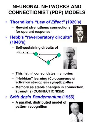
New development: The C.A.T. computer and Fox’s pussycat experiment with it. • The Computer of Average Transients (CAT; “transient” means temporary signal like an ERP) was adapted by Mary Brazier (a Fox mentor at UCLA) for ERP averaging. (The CAT was initially invented to average atomic wave phenomena.) Fox took one with him to Michigan, then Iowa.
WOW! The CAT had 400 bytes (not MB or GB) of memory! • These bytes were in 100 byte segments, so you could have 4 average erps—say to 4 different stimuli—separately averaging in 4 separate channels. (Before the CAT, people would simply superimpose repeated sweeps into a fuzzy mess on a storage oscilloscope.) • The CAT could make one other thing: the post stimulus time histogram (PSTH)
PSTH is a latency distribution, much like a normal bell curve is a grade distribution.
Latency distribution • After, say, 1000 trials, the numbers in each bin in the memory string (called a “buffer”) would contain the number of times in 1000 sweeps that spikes occurred at that particular time. • This is a decent approximation to the probability of a spike at, say, 200 msec (or whatever time bin), post stimulus. If a spike occurs on 500 trials at 200 msec post-stimulus, the probability of a spike at 200 ms = around .5. • So in the PSTH, you have a sequence of spike probabilities for various times following the stimulus. It is indeed a probability distribution, a distribution of firing probabilities at various times(latencies) after the stimulus. The post stimulus time histogram (PSTH) is like any other histogram = distribution.
CAT “software” • To decide whether one of the 4 CAT channels would accumulate average ERPs or PSTHs, the operator pulled the side case of the CAT away and either pulled out a 3 by 5 inch circuit board (for the PSTH) or left it in (for the average ERP, or AERP). • (Some “program!”)
The Fox-O’Brien (1965) cat (the meow type) preparation: • They prepared a cat, under general anesthesia, with scalp retracted, hole drilled in skull, through which a micro-electrode was passed until a single neuron was encountered. Then the cat’s eyes were opened and maintained with lubricant, and a light flashed every 2 seconds.
The Fox-O’Brien (1965) cat (the meow type) preparation • The electrode output was divided into 2 channels, a high pass channel and a low pass channel. • The former was interfaced to the CAT PSTH generator, the latter to an AERP generator:
They took the new CAT out of the box, operated on and set up the CAT connected to the cat… • Then, the went out to dinner, as the lights flashed in the cat’s face, every 2 sec, and the PSTH in one channel, and AERP in the other continued developing.