EXCRETORY (URINARY) SYSTEM
EXCRETORY (URINARY) SYSTEM. I. Urinary system - General information A. Kidneys - semetrically paired organs that form the urine. B. Ureters - carry urine to bladder, one from each kidney. C. Bladder - site where urine is collected and stored until urination.


EXCRETORY (URINARY) SYSTEM
E N D
Presentation Transcript
I. Urinary system - General information • A. Kidneys - semetrically paired organs that form the urine. • B. Ureters - carry urine to bladder, one from each kidney. • C. Bladder - site where urine is collected and stored until urination. • D. Urethra - carries urine from bladder to external environment. http://www.msms.doe.k12.ms.us/biology/anatomy/urinary/urinary.html
II. Kidney • A. General gross external characteristics • 1. Bean shaped => concave on one side, convex on the other • 2. Kidney is surrounded by a dense connective tissue capsule. • 3. An indentation in the concave side is called the hilus - where nerves, blood vessels and lymph vessels enter and leave. • 4. Renal pelvis - expanded end of the ureter that also connects to the hilus. A region where urine from the kidney collects and drains into ureter.
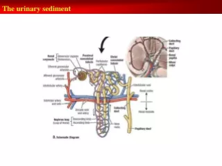
1. Renal cortex contains • (where urine is produced) • a. Upper portions of nephrons • b. Upper portions of the collecting ducts in medullary rays • 2. Renal medulla is organized as • (where urine is collected) • a. pyramid shaped bundles consisting of the thick and thin portions of the loops of Henle, and many collecting ducts that empty into • b. the minor calyces. These connect to the • c. major calyces that empty into the renal pelvis (expanded end of the ureter). B. The Kidney can be divided into a cortex and a medulla.(Has to do with function as well as position)
3. Renal medulla Modified from http://wberesford.hsc.wvu.edu/Kidney.ppt http://www.georgetown.edu/dml/educ/micro/urin/4.htm a. The medulla can be subdivided into structures called the medullary or Malpighian pyramids. b. These pyramids of tissue have there vertex (the pointed end called the papilla) at a minor calyx and their base at the border of the cortex.
http://www.udel.edu/Biology/Wags/histopage/colorpage/cu/cumclhpd.GIFhttp://www.udel.edu/Biology/Wags/histopage/colorpage/cu/cumclhpd.GIF • c. The pyramid tissues consist of • * Collecting ducts that transfer urine from the nephrons in the cortex to the minor calyces • * The thick and thin portions of the loops of Henle. • * capillaries • * Each side of the pyramid that extends toward the cortex is bordered by an interlobular artery
d. Where the medullary pyramid tissue meets the cortex their are large arched blood vessels called arcuate vessels, branches of which extend into the cortex. e. Portions of the medullary tissues called medullary rays (=pars radiata) project into the cortical regions http://wberesford.hsc.wvu.edu/Kidney.ppt http://www.leeds.ac.uk/chb/pcd2130/Img0044.jpg http://anatomy.utmb.edu/microanatomy/kidney/kidney.htm#Kidney Glomerulus http://www.lab.anhb.uwa.edu.au/mb140/
f. The medullary rays subdivide the cortex into regions called cortical labyrinths (=pars convolutae). g. The cortical labyrinths consist of many Bowman's capsules, and proximal and distal convoluted tubules and have a “tortuous” appearance. h. Note that in addition to the cortical areas between the capsule and the bases of the pyramids, cortical tissue also extends between adjacent pyramids. These areas are called the renal columns of Bertin. http://wberesford.hsc.wvu.edu/Kidney.ppt http://www.udel.edu/Biology/Wags/histopage/colorpage/cu/cukcmr.gif http://128.218.123.161/IDS_100/urine/fig1.html
C. The nephron - the nephron is the functional unit of the kidney. There are approximately one million nephrons in each human kidney. They consist of 2 components - • * renal corpuscle • * renal tubules
1. Renal corpuscle - 4 parts- Bowman’s capsule, glomerulus, afferent arteriole, efferent arteriole. • a. Bowman's capsule • * Can be considered to be the expanded and invaginated end of the proximal convoluted tubule. • * Outer surface - parietal layer • **composed of a simple squamous epithelium Renal corpuscle
* Inner surface - visceral layer • ** in intimate contact with the capillaries • ** Composed of cells called podocytes that have many processes • ** These processes (pedicels) intimately surround the capillaries of the glomerulus. • ** The pedicels interdigitate with each other and attach to the basal lamina of the capillary endothelium. http://www.leeds.ac.uk/bms/teaching/histology/histology_term3_frames.htm
** Spaces between the interdigitating pedicels are called slit diaphrgms or filtration slits • ** Filtrate that will form the urine leaves the fenestrated capillaries and enters Bowman's capsule through the filtration slits. • ** The function of the podocytes is probably mechanical. They probably act to prevent the rupture of the glomerular capillaries due to blood pressure and at the same time allow filtration to proceed.
* space between the parietal and visceral layers is called the capsular space - where filtrate collects Renal corpuscle
b. Glomerulus • * Composed of a tuft of tortuous capillary loops that arise from the afferent arteriole and connect to the efferent arteriole. • * The capillary walls are highly fenestrated and completely encircled by a continuous basal lamina (formed from fusion of endothelial cell and podocyte basal laminae, see below). • * This arrangement acts to form a selective filter that will allow certain components of the blood plasma (including excretory wastes) to pass into Bowman's capsule. Renal corpuscle http://www.lab.anhb.uwa.edu.au/mb140/
Renal corpuscle • c. Afferent arteriole • * Typical arteriole except for the portion close to the glomerulus • ** In this area it looses it's internal elastic lamina • ** Smooth muscle cells of tunica media become enlarged and glandular • ** These are thejuxtaglomerular cellsthat secrete the enzymereninthat is involved in the control of blood pressure.
* At the point where the distal tubule is adjacent to the afferent and efferent arterioles of it`s own renal corpuscle, the structure of its epithelium changes (called the juxtaglomerular region). * Cells of the distal tubule wall become more columnar and take on a darker stain. * This region is called the macula densa. * Function of the macula densa -provides "information" concerned with the composition of filtrate in the distal tubule. This “information” in the form of molecular signals, regulates the secretion of renin. http://www.finchcms.edu/anatomy/histology/organology/urinary/o_u_6.html
* Function of renin • ** Converts plasma protein angiotensinogin (synthesized and secreted by the liver hepatocytes) into angiotensin I. • ** The angiotensin I is carried to the lungs by the circulatory system and there it is converted to angiotensin II by an enzyme in lung tissue. • ** Angiotensin II is a powerful vasoconstrictor that causes contraction of smooth muscle in the tunica media of arteries and a resultant increase in systemic blood pressure. • ** The angiotensin II also causes an increase in secretion of the hormone aldosterone by the adrenyl cortex (zona glomerulosa). • ** Aldosterone acts on cells of renal tubules causing increased reabsorption of sodiumand chloride ions from the filtrate. • ** This reabsorption of sodium and its increased concentration in the blood results in additional retension of water and causes further increase in the systemic blood pressure.
d. Efferent arteriole • * Not called a venule because structure is like an arteriole - thicker tunica media than in venule • * Similar to afferent arteriole, but fewer juxtaglomerular cells • * Almost immediately breaks up into two capillary beds that surround the convoluted tubules and the loop of Henle. Renal corpuscle
2. Renal tubule - extends from Bowman's capsule, through cortex, into medulla, back into cortex where it connects with a collecting duct - 3 parts • a. Proximal convoluted tubule • * Filtrate exits Bowman’s capsule through proximal convoluted tubule. • * The tubule wall is composed of a simple cuboidal epithelium with a microvillar brush border along the lumen of the tubule.
* Cells have many mitochondria, a central, spherical nucleus, and a well developed basement membrane. * Lateral walls of these cells interdigitate with each other. * The apical plasmelemma shows very active pinocytosis between microvilli. This is because these cells are responsible for reabsorption of proteineceous molecules from the filtrate. * These cells also reabsorb 75 - 80% of the water and sodium ions in the filtrate, as well as certain sugars and amino acids. http://www.finchcms.edu/anatomy/histology/organology/ urinary/o_u_3.html http://128.218.123.161/IDS_100/urine/fig5.html http://www.meddean.luc.edu/lumen/MedEd/Histo/frames/h_fram16.html http://www.medsch.wisc.edu/anatomy/histo/htm/turibase.htm
b. Loop of Henle • Those near capsule- cortical nephrons (no thin ascending loop of Henle) vs. those near cortical-medullary border - juxtamedullary nephrons (very long loops of Henle with thin descending and ascending portions). • * Each loop has thick and thin segments. • ** Thick segments are similar in structure to the distal convoluted tubule. Also - transition zones: decending thick segment goes from a simple cuboidal to simple squamous epithelium, ascending thick segment goes from a simple squamous to a simple cuboidal epithelium. • ** Thin segment resembles a blood capillary with somewhat thicker walls than normal. • **The loop of Henle further concentrates the urine by the removal of additional water by osmotic diffusion. • **Juxtamedullary loops of Henle extend deep into Malpighian pyramids - responsible for ability to produce hypertonic urine. http://www.finchcms.edu/anatomy/histology/organology/urinary/o_u_4.html
c. Distal convoluted tubule • * Lined by simple cuboidal epithelium. • On your lab slides these cells will look similar to those that line the proximal tubule, however they lack a microvillar brush boarder! • * Cell nucleus usually adjacent to lumenal plasmalemma in fixed material. • * Reabsorption of sodium ions and water (~9% of what’s left). Secretion of potassium and hydrogen ions into lumen - control of acid-base balance. http://www.medsch.wisc.edu/anatomy/histo/htm/turibase.htm http://www.meddean.luc.edu/lumen/MedEd/Histo/frames/h_fram16.html
Identify tubules, etc. http://www.meddean.luc.edu/lumen/MedEd/Histo/frames/h_fram16.html
D. Collecting tubules and ducts • 1. The distal convoluted tubules of the nephrons empty into the collecting tubules. • 2. The collecting tubules extend into the renal medulla and merge to form the large papillary ducts of Bellini that empty into the calyces. • 3. The smaller tubules are lined with simple cuboidal epithelium. As they penetrate deeper into the medulla and approach the papillary ducts, the lining becomes columnar. May even become stratified cuboidal. • 4. The collecting tubules and papillary ducts are regions of additional water re-absorption. They also act to transfer the urine to the calyxes. http://www.finchcms.edu/anatomy/histology/organology/urinary/o_u_5.html
E. Cardiovascular circulation to the kidney • 1. The kidneys receive blood from renal arteries. • 2. These enter the kidney through the connective tissue of the hilus. • 3. Within the hilus these arteries branch to form the interlobar arteries which extend between the medullary pyramids. • 4. As the interlobular arteries reach the juxtamedullary region, they branch to form the arcuate arteries that run parallel to the connective tissue capsule surrounding the kidney at the level of the cortical-medullary junction.
5. Branches from the arcuate arteries extend perpendicular into the cortex and give rise to the afferent arterioles of the glomeruli. 6. The capillaries of the glomerulus re-coalesce to form the efferent arteriole that leaves the glomerulus and then rebranches to form two capillary networks, a. one surrounding the proximal and distal convoluted tubules and b. the other forms a capillary net around the loop of Henle. This second type of capillary network is characteristic of the juxtamedullary nephrons where the capillaries extend deep into the medulla (Malpighian pyramids) P. 360 in text (might be a few pages off)
7. In the case of the juxtamedullary (next to the medulla) nephrons, one branch of the efferent arteriole follows a linear path into the medulla where it breaks up into linear capillaries that loop back toward the cortex where they form venules that will join the arcuate vein. These linear capillaries are called the vasa recta and provide oxygen and nutrients to the tissues of the medulla. http://www.finchcms.edu/anatomy/histology/organology/urinary/o_u_4.html
8. The arcuate veins connect to interlobar veins that extend parallel to their corresponding interlobular arteries. 9. The interlobular veins connect to the renal vein that carries blood out of the kidney. 10. There are also the stellate veins in the peripheral cortex of the kidney that result from the convergence of capillaries in this area. These also empty blood into the arcuate veins.
E. The calyxes, pelvis, ureter, bladder and urethral structure is relatively simple. • F. Mucosa, muscularis and adventitia present. • G. Epithelia of the calyxes, ureter, bladder, and urethra • 1. Calyxes - transitional (2-3 cells thick) • 2. Ureter - transitional (4-5 cells thick) • 3. Bladder - transitional (6-8 cells thick) • 4. Urethra • a. Males • * Prostatic urethra - transitional • * Membranous urethra - pseudostratified or stratified columnar • * Penile urethra - pseudostratified or stratified columnar except at opening in glans where it is stratified squamous • b. Females • * Transitional near bladder, the rest is stratified squamous. There may be a short region of pseudostratified or stratified columnar epithelium between the transitional and squamous zones.