What You Can Do With Our Two-Photon Microscope
200 likes | 611 Vues
What You Can Do With Our Two-Photon Microscope. Genevieve Phillips Fluorescence Microscopy Open House November 12 th , 2012. Our newest piece of equipment. Zeiss LSM510 META Two-Photon Microscope Two external detectors (more sensitive) Coherent Chameleon Ti:Sapphire laser

What You Can Do With Our Two-Photon Microscope
E N D
Presentation Transcript
What You Can Do With OurTwo-Photon Microscope Genevieve Phillips Fluorescence Microscopy Open House November 12th, 2012
Our newest piece of equipment • Zeiss LSM510 META Two-Photon Microscope • Two external detectors (more sensitive) • Coherent Chameleon Ti:Sapphire laser • Tunable wavelength 680-1080nm • Blue, Green, and Red • Located in CRF 226 • Available for training and use
What is two-photon microscopy • A microscopy technique using an IR laser, allowing the researcher to image deeper • Longer wavelength = less scatter • Live or fixed samples • Can acquire z-stacks to create 3D images • Can collect time series data
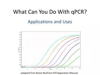
How it works • Pulses of high intensity, IR light enters sample • A fluorescent molecule absorbs two photons simultaneously • only likely in focal volume where light is focused http://micro.magnet.fsu.edu/primer/java/multiphoton/excitationregion/index.html
How it works • The combined energy of the two photons is now enough to excite the fluorophore http://www.microscopyu.com/articles/fluorescence/multiphoton/multiphotonintro.html http://micro.magnet.fsu.edu/primer/java/multiphoton/jablonski/index.html
How it works • Collect all emission • no pinhole • Optical section is created because only fluorescence from focal volume is excited. http://www.microscopyu.com/articles/fluorescence/multiphoton/multiphotonintro.html http://micro.magnet.fsu.edu/primer/java/multiphoton/excitationbleaching/index.html
Differences Between Single-Photon and Two-Photon Microscopy Single-Photon (regular confocal) Two-Photon Two photons of approx double optimal wavelength excites fluorophore Excite fluorescence only in focal volume Photobleaching occurs only in focal volume Collect all emitted light – no pinhole • One photon at optimal wavelength excites fluorophore • Excite fluorescence throughout sample • Photobleaching occurs throughout sample • Use pinhole to only collect emission from focal plane
What is two photon microscopy is good for? • Thick tissue sections • Whole organ • Whole animal (but not on this microscope) Why? • Deep penetration • Less damaging (mostly)
What it is not good for • Thin layer of cultured cells • Some structures, especially aqueous environments, absorb the IR light and produce damaging amounts of heat. Heat damage from IR absorption Image with no heat damage Dr. Diane Lidke
Mara Steinkamp PhD • Spheroids – a large group of cultured cells (20,000 cells) grow into a sphere • Red – RFP – all cells • Green – AF488 – Pertuzumab • Want to know if green penetrates into spheroid. • Used IR wavelength to penetrate deeper. Increased from 100 um to 190 um.
Second Harmonic Generation • Not fluorescence • No labeling of structure • Works only with structures that are highly ordered and non-centrosymmetric, such as collagen fibrils • Most biological structures are optically isotropic – no SHG Dr. Kristina Trujillo
Second Harmonic Generation • High intensity light is required • Two photons interact with a non-centrosymmetric structure • Simultaneously the two photons create one photon with exactly half the wavelength • Specimen does not absorb photon No photobleaching No damage from SHG process http://staff.aist.go.jp/narazaki-aiko/SHG_en.html
Kristina Trujillo PhD • Human Breast Tissue (control) • White – Second Harmonic Generation – Collagen 1 • Blue – DAPI – nuclei • Green – AF 488 – Actin • Far Red – AF 633 – Collagen 4 • Amount of collagen 4 is reduced in tumor adjacent tissue • Looking at the interaction of Collagen 1 and 4
New components coming soon • 25x multi-immersion objective • 40x water objective • Incubation chamberwith perfusion and objective heater
Other Possible Applications • Use IR laser to excite DAPI – META alternative with green, red, far red confocal • Live cell single-photon, regular confocal imaging – META alternative (once we get objective heater and chamber)
Thank You!!! Questions??