Glomerulopathies vs. Glomerulonephritis
Glomerulopathies vs. Glomerulonephritis. Glomerular disease includes glomerulonephritis, i.e. inflammation of the glomeruli and glomerulopathies when there is no evidence of inflammation. Glomerulonephritis is a subset of glomerulopathies. CLASSIFICATION OF GLOMERULOPATHIES.

Glomerulopathies vs. Glomerulonephritis
E N D
Presentation Transcript
Glomerulopathies vs. Glomerulonephritis • Glomerular disease includes glomerulonephritis, i.e. inflammation of the glomeruli and glomerulopathies when there is no evidence of inflammation. • Glomerulonephritis is a subset of glomerulopathies
CLASSIFICATION OF GLOMERULOPATHIES • Nephrotic syndrome. • Acute glomerulonephritis (acute nephritic syndrome). • Rapidly progressive glomerulonephritis. • Asymptomatic urinary abnormality (haematuria, proteinuria or both).
Glomerular disease • Primary – confined to the kidney • Secondary – due to a systemic disease
Possible Clinical Manifestations • Proteinuria – asymptomatic • Haematuria – asymptomatic • Hypertension • Nephrotic syndrome • Nephritic syndrome • Acute renal failure • Rapidly progressive renal failure • End stage renal failure 4
Glomerulonephritis • Presence of glomerular disease as opposed to tubulointersititial or vascular disease is suspected from history • Haematuria (especially dysmorphic red cells) • Red cell casts • Lipiduria (glomerular permeability must be increased to allow the filtration of large lipoproteins) • Proteinuria (may be in nephrotic range of >3.5 g/24hours) 5
Pathology Immune complex disease • Complement-leukocyte- mediated mechanism • Activation of the complement pathway Recruitment of neutrophils and monocytes Complement- dependent C₅- C₉ (MAC) • Epithelial cell detachment. • (+) epithelial & mesangial cells to secrete damaging chemical mediators. • Upregulates TGF receptors on epithelial cells, excessive synthesis of extracellular matrix which leads to GBM thickening • Neutrophils: • Protease GBM degradation • O₂ free readicals cell damage • AA metabolites ↓ GFR
Glomerular Disease • Named according to • etiology • microscopic findings • clinical syndrome • Most common clinical presentations • acute nephritic syndrome • nephrotic syndrome • Most common cause is autoimmune
Autoimmune Reactions Some progress as either focal segmental glomerulosclerosis or tubulointerstitial nephritis
Poststreptococcal Glomerulonephritis • Autoimmune injury initiated by beta-hemolytic streptococcus • aka acute proliferative glomerulonephritis • Presents as acute nephritic syndrome • hematuria • HT • increased urea & creatinine • low urine output • edema • Antibodies produced by strep throat deposit in glomerulus • Most fully recover but about 10% evolve into rapidly progressive glomerulonephritis
Rapidly Progressive Glomerulonephritis • Unknown causes or secondary to poststreptococcal glomerulonephritis • Autoimmune • aka crescentric glomerulonephritis • Some present as acute nephritic syndrome & others as renal failure • Caused by deposition of An-Ab complexes • All but a few progress to renal failure
Membranous Glomerulonephritis • Autoimmune • Most common cause of nephrotic syndrome in adults • About 10% proceed to renal failure within 10 yrs, 25% recover completely, most progress slowly with proteinuria, HTN, loss of renal function
Chronic Glomerulonephritis • Incidental discovery of occult proteinuria or HTN • Usually presents as chronic renal failure or occult proteinuria • Glomerulus has scar tissue • Dialysis & transplant
Secondary Glomerulonephritis • Diabetes most common cause • most common cause of renal failure • glycoproteins deposit in basement membrane • Vascular disease • atherosclerosis • HTN • vascultitis
Characteristics of common glomerular diseases at presentation
Definition • Acute glomerulonephritis is the inflammation of the glomeruli which causes the kidneys to malfunction • It is also called Acute Nephritis, Glomerulonephritis and Post-Streptococcal Glomerulonephritis • Predominantly affects children from ages 2 to 12 • Incubation period is 2 to 3 weeks
Etiology • Infectious • Streptococcal • Nonstreptococcal postinfectious glomerulonephritis • Bacterial • Viral • Parasitic • Noninfectious • Multisystem systemic diseases • Primary glomerular diseases
Pathogenesis Previously M-protein of the organism was felt to be responsible for PSGN. Recently, nephritis-associated streptococcal cationic protease and its zymogen precursor (NAPR) has been identified as a glyceraldehyde-3-phosphate dehydrogenase that functions as a plasmin(ogen) receptor. Antibody levels to NAPR are elevated in streptococcal infections (of group A, C, and G) associated with glomerulonephritis, but are not elevated in streptococcal infections without glomerulonephritis, where as anti-streptolysin-O titers are elevated in both circumstances.
Pathology of Acute glomerulunephritis Diffuse proliferative GN (PGN) • proliferation of cells within the glomeruli, accompanied by leukocyte filtrate • typical features of immune complex disease : • - hypocomplimentemia • - granular deposits of IgG & complement on GBM • Implicated antigens seem to be endostreptosin and nephritis – plasmin- binding ptn
General Symptoms • Fever • Headache • Malaise • Anorexia • Nausea and vomiting • High blood pressure • Pallor due to edema and/or anemia • Confusion • Lethargy • Loss of muscle tissue • Enlargement of the liver
Signs and Symptoms • Hematuria: dark brown or smoky urine • Oliguria: urine output is < 400 ml/day • Edema: starts in the eye lids and face then the lower and upper limbs then becomes generalized; may be migratory • Hypertension: usually mild to moderate
Clinical syndromes • urinary (haematuria, proteinuria), • nephritic (edemas, hypertension, gross haematuria, proteinuria), • nephrotic (edemas, proteinuria, hypoproteinemia, hypercholesterolemia), • mixed.
CLINICAL FEATURES • Abrupt onset of: • glomerular haematuria (RBC casts or dysmorphic RBC). • non-nephrotic range proteinuria (<2 g in 24 hrs). • oedema (periorbital, sacral). • hypertension. • transient renal impairment (oliguria, uraemia).
INVESTIGATIONS • Base line measurements: • ↑ Urea • ↑ Creatinine • Urinalysis (MSU): • a) Urine microscopy (red cell cast) • b) proteinuria
Complications • Hypertensive encephalopathy, heart failure and acute pulmonary edema may occur in severe cases • Acute renal necrosis due to injury of capillary or capillary thrombosis
Prevention • proper hygiene • prompt medical assessment for necessary antibiotic therapy should be sought when infection is suspected • prophylactic immunizations
Treatment Treat the underlying infections when acute GN is associated with chronic infections. • Antimicrobial therapy • Antibiotics (eg, penicillin) are used to control local symptoms and to prevent spread of infection to close contacts. • Antimicrobial therapy does not appear to prevent the development of GN, except if given within the first 36 hours. • Loop diuretic therapy • Loop diuretics may be required in patients who are edematous and hypertensive in order to remove excess fluid and to correct hypertension. • Relieves edema and controls volume, thereby helping to control volume-related elevation in BP. • Vasodilator drugs (eg, nitroprusside, nifedipine, hydralazine, diazoxide) may be used if severe hypertension or encephalopathy is present • Diet: • Sodium and fluid restriction • Protein restriction for azotemic patients • Activity:Recommend bed rest until signs of glomerular inflammation and circulatory congestion subside.
Management & Prognosis Post streptococcal GN - Has a GOOD prognosis. - Supportive measures until spontaneous recovery. - Control HT. - Fluid balance. - Oliguric with fluid overload. - GN complicating SLE or systemic vasculitides: immunosuppression with prednisolone, cyclophosphamide or azathioprine/MMF.
Chronic glomerulonephritis The condition is characterized by irreversible and progressive glomerular and tubulointerstitial fibrosis, ultimately leading to a reduction in the glomerular filtration rate (GFR) and retention of uremic toxins. If disease progression is not halted with therapy, the net result is chronic kidney disease (CKD), end-stage renal disease (ESRD), and cardiovascular disease. The diagnosis of CKD can be made without knowledge of the specific cause.
Etiology Nearly all forms of acute glomerulonephritis have a tendency to progress to chronic glomerulonephritis. The progression from acute glomerulonephritis to chronic glomerulonephritis is variable. Whereas complete recovery of renal function is the rule for patients with poststreptococcal glomerulonephritis, several other glomerulonephritides, such as immunoglobulin A (IgA) nephropathy, often have a relatively benign course and many do not progress to ESRD.
Pathogenesis Reduction in nephron mass from the initial injury reduces the GFR. This reduction leads to hypertrophy and hyperfiltration of the remaining nephrons and to the initiation of intraglomerular hypertension. These changes occur in order to increase the GFR of the remaining nephrons, thus minimizing the functional consequences of nephron loss. The changes, however, are ultimately detrimental because they lead to glomerulosclerosis and further nephron loss.
Histologic Findings In early stages, the glomeruli may still show some evidence of the primary disease. In advanced stages, the glomeruli are hyalinized and obsolescent. The tubules are disrupted and atrophic, and marked interstitial fibrosis and arterial and arteriolar sclerosis occur.
Minimal-Change Disease fusion of podocytes on electron microscopy
Focal segmental glomerulosclerosis Segmental areas of glomerular sclerosis, hyalinization of glomerular capillaries
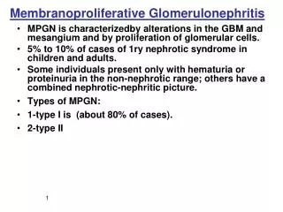
Mesangiocapillary GN large glomeruli with mesangial proliferation and ‘double’ BM. 2 histological types: type I (subendothelial deposits) type II (intramembranous deposits)
Membranous nephropathy thickened BM, IF +ve for IgG & C3 and subepithelial deposits on EM
Mesangial proliferativeGN Hypercellularity, mesangial proliferation, inflammatory cell infiltrate, positive IF for IgG and C3 and subepithelial deposits on EM.
Clinical Manifestations • Uremia-specific findings • Edemas • Hypertension • Jugular venous distension (if severe volume overload is present) • Pulmonary rales (if pulmonary edema is present) • Pericardial friction rub in pericarditis • Tenderness in the epigastric region or blood in the stool (possible indicators for uremic gastritis or enteropathy) • Decreased sensation and asterixis (indicators for advanced uremia)
Clinical variants • Latent (changes in urine) • Hypertensive (increased blood pressure) • Hematuric • Nephrotic (edemas, proteinuria, hypoproteinemia, hypercholesterolemia), • Mixed
Lab Studies • Urinalysis • Urinary protein excretion • Serum chemistry • Serum creatinine and urea nitrogen levels are elevated. • Impaired excretion of potassium, free water, and acid results in hyperkalemia, hyponatremia, and low serum bicarbonate levels, respectively. • Impaired vitamin D-3 production results in hypocalcemia, hyperphosphatemia, and high levels of parathyroid hormone. • Low serum albumin levels may be present if uremia interferes with nutrition or if the patient is nephrotic.
ImagingStudies • Renal ultrasonogram • Obtain a renal ultrasonogram to determine renal size, to assess for the presence of both kidneys, and to exclude structural lesions that may be responsible for azotemia. • Small kidneys often indicate an irreversible process. • Kidney biopsy
Treatment • The target pressure for patients with proteinuria greater than 1 g/d is less than 125/75 mm Hg; for patients with proteinuria less than 1 g/d, the target pressure is less than 130/80 mm Hg. • Angiotensin-converting enzyme inhibitors (ACEIs) • angiotensin II receptor blockers (ARBs) • combination therapy with ACEIs and ARBs. • Diuretics are often required because of decreased free-water clearance, and high doses may be required to control edema and hypertension when the GFR falls to less than 25 mL/min. • Beta-blockers, calcium channel blockers, central alpha-2 agonists (eg, clonidine), alpha-1 antagonists, and direct vasodilators (eg, minoxidil, nitrates) may be used to achieve the target pressure.
Treatment • Renal osteodystrophy can be managed early by replacing vitamin D and by administering phosphate binders. • Seek and treat nonuremic causes of anemia, such as iron deficiency, before instituting therapy with erythropoietin. • Discuss options for renal replacement therapy (eg, hemodialysis, peritoneal dialysis, renal transplantation). • Treat hyperlipidemia (if present) • Expose patients to educational programs for early rehabilitation from dialysis or transplantation.
Treatment Minimal change glomerulonephritis (MCGN) Corticosteroids induce remission in >90% of children and 80% of adults (slower response). Indications for immunosuppression: (cyclophosphamide, ciclosporin (=cylosporin)): early/ frequent relapses; steroid SEs/dependence. Prognosis: 1% progress to ESRF.
Treatment Focal segmental glomerulosclerosis Poor response to corticosteroids (10–30%). Cyclophosphamide or ciclosporin (=cylosporin) may be used in steroid-resistant cases. Prognosis: 30–50% progress to ESRF.
Treatment Mesangiocapillary GN Treatment: None is of proven benefit. Prognosis: 50% develop ESRF.
Treatment Membranous nephropathy If renal function deteriorates, consider corticosteroids and chlorambucil. Prognosis: Untreated, 15% complete remission, 9% ESRF at 2–5yrs and 41% at 15yrs.
Treatment Mesangial proliferative GN Antibiotics, diuretics, and antihypertensives as necessary. Dialysis is rarely required. Prognosis: Good.