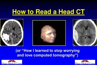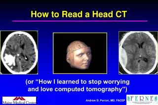
How to Read a Head CT
How to Read a Head CT. Dr Mohamed El Safwany . MD. Intended learning outcome. The student should learn at the end of this lecture interpretation of CT Brain. Head CT.

How to Read a Head CT
E N D
Presentation Transcript
How to Read a Head CT Dr Mohamed El Safwany. MD.
Intended learning outcome The student should learn at the end of this lecture interpretation of CT Brain.
Head CT • Has assumed a critical role in the daily practice of Emergency Medicine for evaluating intracranial emergencies. (e.g. Trauma, Stroke, SAH, ICH). • Most practitioners have limited experience with interpretation. • In many situations, the Emergency Physician must initially interpret and act on the CT without specialist assistance.
Head CT “Blood Can Be Very Bad”
Blood Can Be Very Bad • Blood • Cisterns • Brain • Ventricles • Bone
Blood Can Be Very Bad • Blood • Cisterns • Brain • Ventricles • Bone
Blood Can Be Very Bad • Blood • Cisterns • Brain • Ventricles • Bone
Blood Can Be Very Bad • Blood • Cisterns • Brain • Ventricles • Bone
Blood Can Be Very Bad • Blood • Cisterns • Brain • Ventricles • Bone
CT Scan Basics • A CT image is a computer-generated picture based on multiple x-ray exposures taken around the periphery of the subject. • X-rays are passed through the subject, and a scanning device measures the transmitted radiation. • The denser the object, the more the beam is attenuated, and hence fewer x-rays make it to the sensor.
CT Scan Basics • The denser the object, the whiter it is on CT • Bone is most dense = + 1000 Hounsfield U. • Air is the least dense = - 1000H Hounsfield U.
CT Scan Basics: Windowing Focuses the spectrum of gray-scale used on a particular image.
Posterior Fossa • Brainstem • Cerebellum • Skull Base • Clinoids • Petrosal bone • Sphenoid bone • Sella turcica • Sinuses
2nd Key Level Sagittal View 2nd Key Level Circummesencephalic Cistern
3rd Key Level Sagittal View Circummesencephalic Cistern
CSF Production • Produced in choroid plexus in the lateral ventricles Foramen of Monroe IIIrd Ventricle Acqueduct of Sylvius IVth Ventricle Lushka/Magendie • 0.5-1 cc/min • Adult CSF volume is approx. 150 cc’s. • Adult CSF production is approx. 500-700 cc’s per day.
1 day 1 year 2 years Andrew D. Perron, MD, FACEP 25
B is for Blood • 1st decision: Is blood present? • 2nd decision: If so, where is it? • 3rd decision: If so, what effect is it having?
B is for Blood • Acute blood is bright white on CT (once it clots). • Blood becomes hypodense at approximately 2 weeks. • Blood becomes isodense at approximately 1 week.
B is for Blood • Acute blood is bright white on CT (once it clots). • Blood becomes hypodense at approximately 2 weeks. • Blood becomes isodense at approximately 1 week.
B is for Blood • Acute blood is bright white on CT (once it clots). • Blood becomes hypodense at approximately 2 weeks. • Blood becomes isodense at approximately 1 week.
Subdural Hematoma • Typically falx or sickle-shaped. • Crosses sutures, but does not cross midline. • Acute subdural is a marker for severe head injury. (Mortality approaches 80%) • Chronic subdural usually slow venous bleed and well tolerated.
Subarachnoid Hemorrhage • Blood in the cisterns/cortical gyral surface • Aneurysms responsible for 75-80% of SAH • AVM’s responsible for 4-5% • Vasculitis accounts for small proportion (<1%) • No cause is found in 10-15% • 20% will have associated acute hydrocephalus
CT Scan 34
CT Scan 35
CT Scan 37
C is for CISTERNS (Blood Can Be Very Bad) • 4 key cisterns • Circummesencephalic • Suprasellar • Quadrigeminal • Sylvian Circummesencephalic
Cisterns • 2 Key questions to answer regarding cisterns: • Is there blood? • Are the cisterns open?
B is for BRAIN (Blood Can Be Very Bad)
Tumor 44
Atrophy 45
Abscess 46
Mass Effect 48