Download
1 / 7
170 likes | 826 Vues
NYSED Living Environment Part D Diffusion Through a Membrane Lab Review. Mrs. Koch. Diffusion and Osmosis. Designed to help you understand the concepts of Diffusion and Osmosis and how these cell processes effect the cell;

E N D
NYSED Living Environment Part D Diffusion Through a Membrane Lab Review Mrs. Koch
Diffusion and Osmosis • Designed to help you understand the concepts of Diffusion and Osmosis and how these cell processes effect the cell; • Define: diffusion, osmosis, saline, selectively permeable, molecule size;
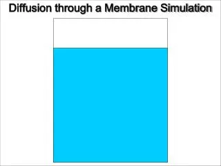
Part 1: Diffusion • Diffusion: movement of molecules from an area of high concentration to an area of low concentration along the concentration gradient. • Example is when you put your Lugol’s (iodine) solution into the water and the water began to turn the “amber/tea” color. After diffusion occurs… Before diffusion occurs…
Part 2: Create a “cell” • Soak 10 inches of dialysis tubing; • Tie knot in one end; • Put 10mL of glucose solution in and 20mL of starch solution in; • Pinch/clamp closed and put into cellular environment (Lugol’s (iodine) and water) for 15 minutes; • Observe the changes and infer what happened Wait about 15 minutes and observe. Insert into “cellular environment”
After 15 minutes, observe… • What happened to the glucose in the “cell”? …the starch in the “cell”? …the Lugol’s iodine outside the “cell”? Why? S S G I G G G I I G I G I S S Iodine solution (I) I I I Glucose solution (G) Starch solution (S) At the beginning… After 15 minutes…
And now, the part that makes you cry (ok, not really, but the “Red Onion” part of the lab)… • Prepare a wet mount slide of the inner epidermis of a red onion section; • Observe the red onion and draw what you see; • Add a couple of drops of saline (salt) solution to the epidermis. Wait 5 minutes; • Observe under microscope again, note any changes; • Add freshwater to the slide, wait 5 minutes, observe changes again.
Red Onion Plasmolysis Observation • Before and after observations of red onion epidermis under the microscope (400X) Red onion under in hypertonic (salt) solution. Note cell membrane has “withdrawn” and the cytoplasm has lost water to the salty environment, making it appear smaller and darker. Red onion under in isotonic (normal) solution. Note cell membrane and cytoplasm almost completely “fill” the boundary of the cell wall.