Download

1 / 37
370 likes | 511 Vues
Quantitative Assessment of Tissue-based IHC Biomarkers. Next Generation Pharmaceutical Summit David Young 7 Apr 09. Digital Pathology. Digital Pathology – Research and Clinical Possibilities Quantitative Digital pathology IHC – Traditional evaluation vs Image analysis

E N D
Quantitative Assessment of Tissue-based IHC Biomarkers Next Generation Pharmaceutical Summit David Young 7 Apr 09
Digital Pathology • Digital Pathology – Research and Clinical Possibilities • Quantitative Digital pathology • IHC – Traditional evaluation vs Image analysis • Tools not limited to pathologists
Digital Pathology • Digital Pathology – Research and Clinical Possibilities • Archival of pathology specimens • Diagnosis • Digital slide conferencing • Consultation • Help from Development Teams • putting the power in the hands of the people who know it best
Quantitative Digital Pathology • Pathologist opinions • Good enough for government work, or • Close, but no cigar • X number of pathologists = Y number of results • Diagnoses • IHC analysis • subjective; based on familiarity of tissue and experience
Immunohistochemistry Analyses and Quantitative Digital Pathology • Not an exact science • Basis of many aspects of drug development and drug selection
Biomarker Scoring Consensus • Clark (2006) – ‘there is no consensus in the literature about how to summarize these scoring assessments into a single determination of EGFR protein expression status as EGFR positive or EGFR negative.’ • ‘Evaluation of the clinical significance of EGFR expression by IHC has been complicated by the use of different antibodies, different scoring systems, and different clinical endpoints.’ Clark, et al: J Thorac Oncol 2006
Importance of Standardized Scoring • Prevalence and tumor surveillance • Prognostic factors • Predictive factors • Comparing study results from a recognized baseline of analysis
IHC Scoring Concordance – Pathologists Variability Concordance Total scoring = 78% Cut point <100 = 92% Concordance Total scoring = 75% Cut point <100 = 100%
Pathologist Variation Pathologist 1 Scores: Y = 0.96X + 3.21 R = 0.987 Pathologist 2 Scores: Y = 0.97X -2.72 R = 0.974 Legend: Red – Pathologist 1 Blue – Pathologist 2
Image Analysis– Lessens Subjectivity of Scoring Quantify: Size (area) Positive cells Negative cells Intensity levels
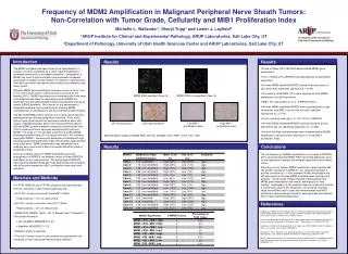
Tissue-based Biomarkers – Case Study • E-Cadherin • Marker of epithelial phenotype • Associated with cell-to-cell adhesion • Membrane protein • Vimentin • Marker of mesenchymal phenotype • Associated with cellular skeleton • Cytoplasmic protein
Experimental Xenograft model H&E E-cad Vim
Traditional IHC Score (H-Score) 1% 10% 30% 100% 0 75% Proportion Score (PS) 0 – 100% Intensity Score (IS) 0 = negative 1 = weak 2 = intermed 3 = strong Score range: 0-300
Factors Affecting IHC Analysis – Not Just the Pathologist • Tumor acquisition (pre-analytical factors) • Tumor size • Tumor type (Tumor tissue and host response) • Antibodies • Processing factors • Individual variation in evaluation
Pancreas – Xenograft 1 H&E E-cad Vim
Summary – What have we learned so far? • Selection of site for IHC evaluation is important; may or may not be reflective of whole tumor • Tumor heterogeneity affects tissue-based biomarker assessment and analysis • IA correlates well with traditional IHC scoring methods. • Validation removes pathologists scoring variability • ‘Tweaking’ of algorithms required prior to universal deployment