Download
1 / 87
880 likes | 1.02k Vues
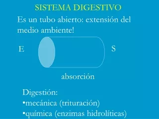
SISTEMA DIGESTIVO. Es un tubo abierto: extensión del medio ambiente!. S. E. absorción. Digestión: mecánica (trituración) química (enzimas hidrolíticas). EL TRACTO DIGESTIVO. Anatomía del sistema digestivo. Digestive tract Alimentary tract or canal GI tract Accessory organs

E N D
SISTEMA DIGESTIVO Es un tubo abierto: extensión del medio ambiente! S E absorción • Digestión: • mecánica (trituración) • química (enzimas hidrolíticas)
Anatomía del sistema digestivo • Digestive tract • Alimentary tract or canal • GI tract • Accessory organs • Primarily glands • Regions • Mouth or oral cavity • Pharynx • Esophagus • Stomach • Small intestine • Large intestine • Anus
Oral Cavity • Mouth or oral cavity • Vestibule: Space between lips or cheeks and alveolar processes • Oral cavity proper • Lips (labia) and cheeks • Palate: Oral cavity roof • Hard and soft • Palatine tonsils • Tongue: Involved in speech, taste, mastication, swallowing
Teeth • Two sets • Primary, deciduous, milk: Childhood • Permanent or secondary: Adult (32) • Types • Incisors, canine, premolar and molars
Salivary Glands • Produce saliva • Prevents bacterial infection • Lubrication • Contains salivary amylase • Breaks down starch • Three pairs • Parotid: Largest • Submandibular • Sublingual: Smallest
SALIVARY SECRETIONS1. Serous secretion that contains ptyalin, which is an enzyme for digesting starches. 2. Mucous secretion that contains mucin for lubricating and for surface protective purposes. 3. Saliva also contains IgA antibodies and lysozyme, which help to destroy any microorganisms in the oral cavity.
Swallowing reflex: Soft Palate & Esophagus Figure 21-13: The swallowing reflex
Deglutition (Swallowing) • Three phases • Voluntary • Bolus of food moved by tongue from oral cavity to pharynx • Pharyngeal Reflex: Upper esophageal sphincter relaxes, elevated pharynx opens the esophagus, food pushed into esophagus • Esophageal • Reflex: Epiglottis is tipped posteriorly, larynx elevated to prevent food from passing into larynx
Peritoneum and Mesenteries • Peritoneum • Visceral: Covers organs • Parietal: Covers interior surface of body wall • Retroperitoneal: Behind peritoneum as kidneys, pancreas, duodenum • Mesenteries • Routes which vessels and nerves pass from body wall to organs • Greater omentum • Lesser omentum
Pharynx Nasopharynx Oropharynx: Transmits food normally Laryngopharynx: Transmits food normally Esophagus Transports food from pharynx to stomach Passes through esophageal hiatus (opening) of diaphragm and ends at stomach Hiatal hernia Sphincters Upper Lower Pharynx and Esophagus
Functions • Ingestion: Introduction of food into stomach • Mastication: Chewing • Propulsion • Deglutition: Swallowing • Peristalsis: Moves material through digestive tract
Stomach Anatomy: • Openings • Gastroesophageal: To esophagus • Pyloric: To duodenum • Regions • Cardiac • Fundus • Body • Pyloric
Stomach Histology: • Layers • Serosa or visceral peritoneum: Outermost • Muscularis: Three layers • Outer longitudinal • Middle circular • Inner oblique • Submucosa • Mucosa
Stomach Histology • Rugae: Folds in stomach when empty • Gastric pits: Openings for gastric glands • Contain cells • Surface mucous: Mucus • Mucous neck: Mucus • Parietal: Hydrochloric acid and intrinsic factor • Chief: Pepsinogen • Endocrine: Regulatory hormones
Gastric Phase: The Stomach Figure 21-15: The mucus-bicarbonate barrier of the gastric mucosa
Small Intestine • Site of greatest amount of digestion and absorption • Divisions • Duodenum • Jejunum • Ileum: Peyer’s patches or lymph nodules • Modifications • Circular folds or plicae circulares, villi, lacteal, microvilli • Cells of mucosa • Absorptive, goblet, granular, endocrine
Small Intestine Secretions • Mucus • Protects against digestive enzymes and stomach acids • Digestive enzymes • Disaccharidases: Break down disaccharides to monosaccharides • Peptidases: Hydrolyze peptide bonds • Nucleases: Break down nucleic acids • Duodenal glands • Stimulated by vagus nerve, secretin, chemical or tactile irritation of duodenal mucosa
Large Intestine, H2O Absorption & Defecation Figure 21-27: Anatomy of the large intestine
Large Intestine Digestion & Absorption • Bacterial fermentation: Vit. K , lactate & butyrate • Water and electrolyte secretion &/or absorption Figure 21-28: NaCl reabsorption by colonocytes
Intestinal Phase: Large Intestine Digestion & Absorption Figure 21-29: NaCl secretion by colonic crypt cells
Phases of Digestion: Overview Figure 21-11: Overview of functions in different regions of the digestive system
Regulating Digestion: CNS and Enteric Nervous System (ENS) Figure 21-9: The enteric nervous system
Cephalic and Oral Phases of Digestion • Cephalic: anticipation of food • CNS ANS long reflex • Enteric cells short reflex • GI motility • GI secretions • Mouth: starts digestion • Grind, mix & liquefy • Saliva: water, enzymes, mucus & lysozyme
Cephalic and Oral Phases of Digestion Figure 21-12: Long and short reflexes in the stomach
Liver • Lobes • Major: Left and right • Minor: Caudate and quadrate • Ducts • Common hepatic • Cystic • From gallbladder • Common bile • Joins pancreatic duct at hepatopancreatic ampulla
Functions of the Liver • Bile production • Salts emulsify fats, contain pigments as bilirubin • Storage • Glycogen, fat, vitamins, copper and iron • Nutrient interconversion • Detoxification • Hepatocytes remove ammonia and convert to urea • Phagocytosis • Kupffer cells phagocytize worn-out and dying red and white blood cells, some bacteria • Synthesis • Albumins, fibrinogen, globulins, heparin, clotting factors
Gallbladder • Bile is stored and concentrated • Stimulated by cholecystokinin and vegal stimulation • Dumps into small intestine • Production of gallstones possible • Drastic dieting with rapid weight loss
Anatomy Endocrine Pancreatic islets produce insulin and glucagon Exocrine Acini produce digestive enzymes Regions: Head, body, tail Secretions Pancreatic juice (exocrine) Trypsin Chymotrypsin Carboxypeptidase Pancreatic amylase Pancreatic lipases Enzymes that reduce DNA and ribonucleic acid Pancreas