Spatial Neglect and Attention Networks
170 likes | 436 Vues
Spatial Neglect and Attention Networks. Stephanie Regan Antoinette Sellers Alyssa Nolde Hannah Stolarczyk. 7. The Core Spatial Deficit and Our Current Understanding. Egocentric Bias

Spatial Neglect and Attention Networks
E N D
Presentation Transcript
Spatial Neglect and Attention Networks Stephanie Regan Antoinette Sellers Alyssa Nolde Hannah Stolarczyk
7. The Core Spatial Deficit and Our Current Understanding • Egocentric Bias • Spatial neglect: represents a deficit of spatial attention and stimulus saliency that is mapped in the egocentric reference frame
7. Continued • Factor Analytic Studies • Have isolated at least one factor associated with impairments in attending, search, and/or responding to targets in contralateral space • Inconclusive in terms of other factors • Classifying Patients • Based on performance on different behavioral tasks that are thought to isolate different subtypes • Ex. Perceptual/ attention VS. motor/intention • Not very consistent or relevant to recovery rates
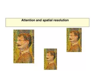
What are measures of contrast sensitivity and segmentation, are they impaired, and why do the authors discuss them? There is an abnormal early visual mechanism, indicated by normal measure of contrast sensitivity and image segmentation based on low-level features in the neglected visual field and responses in occipital cortex based on visually evoked potentials and functional magnetic resonance imaging.
What are measures of contrast sensitivity and segmentation, are they impaired, and why do the authors discuss them? • The author discuss them because… • Almost all patients with spatial neglect form a lateralized bias in visual information processing. • Sensitivity and responsiveness to behaviorally relevant stimuli generally improve as one moves from the contralesional location to the ipsilesional location along the spatial gradient.
What are measures of contrast sensitivity and segmentation, are they impaired, and why do the authors discuss them? • Contrasting the abnormal early visual mechanism, the saliency of object in the neglected field is impaired. • Using task relevant or task irrelevant stimuli, a study found that both stimuli produced an increased probablity of eye movement along the spatial gradient from contalesional to ipsilesional locations. • This suggest that automatic and goal driven components of spatial attention are equally affected.
Explain Figure 1 and the text that discusses it. • Panel A (Bays et al.) • The 1st image shows the difference between ‘target’ and ‘probe’ conditions for letters in arrays. • The 2ndimage shows that targets (red) and probes (blue) produced more saccades in the ipisilesional position than the contralesional position • This provides evidence for the ipsilesional bias for automatic and goal-directed orienting in patients with spatial neglect • The third image shows that lesions are localized or situated in the temporoparietal junction
Explain Figure 1 and the text that discusses it. • Panel B (Fruhmann Berger et al.) • The top graph shows that patients with neglect show an ipsilesional gaze bias while target searching (blue) • Control patients show no gaze bias (blue)
Explain Figure 1 and the text that discusses it. • Panel C (Medina et al.) • The first image displays ipsilesional bias within an egocentric reference frame (viewer’s point of view and midline)
What other functions besides attention or salience are impacted in neglect? Tell us about the research into this issue and what it’s shown. • Proposed that neglect is a deficit in forming, storing, or manipulating the left side of mental images or information in VSTM, termed representational neglect. • Other aspects that could be included in neglect are perceptual cognitive functions such as, visuospatial short-term memory and spatial cognition
11. Damage in which regions can contribute to neglect? Is it necessary that lesions all happen in a specific location, or not? • Parietal Cortex (specifically IPL) • (TOP) • Superior Temporal Gyrus (STG), Inferior Frontal Gyrus (IFG), Anterior Insula, Middle Frontal Gyrus (MFG) • (BOTTOM)
11. Continued • White matter fiber damage can cause a disconnect from the frontal, temporal, and parietal cortex • Damage to the subcortical nuclei: • pulivinar, caudate, putamen • causes cortical hypoactivation of regions associated with neglect
11. Continued • Some lesion sites are more likely than others to produce neglect • Lesions do not always have to be in a specific location • The big question: • Why is it that neglect of the left field can be caused by many different lesions in the right hemisphere?
Figure 2 • Anatomical regions associated with neglect • Shown by lesion-symptom mapping • Lesion overlap in patients diagnosed with neglect • Comparison of patients with severe neglect and no neglect These are quite similar and emphasize ventral regions in IPL
Figure 2 • Attention networks determined by resting state functional connectivity • Yellow-Orange: regions with strong positive temporal correlation of activity at rest • Anatomical distributions in section “a” match distribution of ventral but not dorsal • Blue: regions negatively correlated with dorsal network, corresponds to default network
Figure 2 • Blue and green: regions of maximal damage in neglect patients • White matter tracts corresponding to arcuate fasciculus (AF) and superior longitudinal fasciculus (SLF) II and III