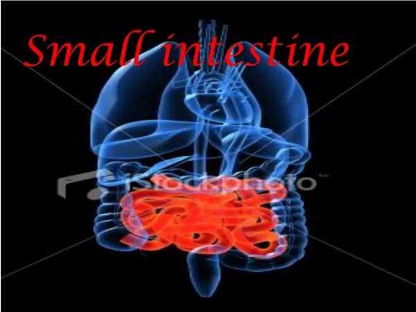
Understanding Small Intestine Anatomy
140 likes | 294 Vues
Explore the microscopic structure and functions of the three regions of the small intestine, including the roles of villi, crypts, and cell types. Learn about surface area enhancement mechanisms and regional differences.

Understanding Small Intestine Anatomy
E N D
Presentation Transcript
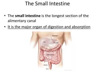
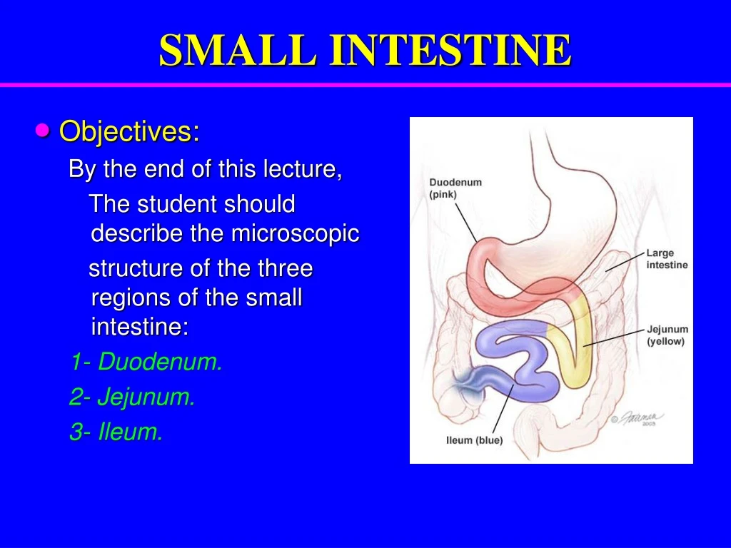
SMALL INTESTINE • Objectives: By the end of this lecture, The student should describe the microscopic structure of the three regions of the small intestine: 1- Duodenum. 2- Jejunum. 3- Ileum.
SMALL INTESTINE • To increase surface area the mucosa has: • Plicae circulares. • Villi. • Intestinal crypts (crypts of Lieberkühn). • Microvilli (Brush border).
Duodenum • Mucosa: Shows villi and crypts. A- Epithelium:simple columnar epithelium with goblet cells. B- Lamina propria: C.T. C- Muscularismucosae: 2 layers of smooth muscle cells.
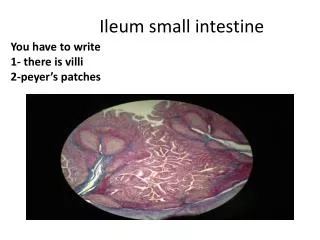
Intestinal villi • Each Villus is a finger-like projection of small intestinal mucosa and it is formed of: I- Central core of loose C.T. containing: • Lymphocytes. • Fibroblasts. • Smooth muscle cells. • Capillary loops. • Lacteal (blindly ending lymphatic channels). II- Villus-covering epithelium.
Cells Covering the Villi 1- Surface columnar absorptive cells: They have brush border (microvilli). They are covered with thick glycocalx that has digestive enzymes. They have Junction complex (tight, adhering and desmosome junctions). 2- Goblet cells: Increase toward the ileum. 3- Enteroendocrine (EE) cells (DNES cells).
Intestinal Glands (Crypts) • Simple tubular glands that open between villi. • Composed of 5 cell types: 1. Columnar absorptive cells. • Goblet cells: secrete mucus. • Enteroendocrine (EE) (DNES) cells: secrete hormones. 4.Paneth cells: secrete Lysozyme (antibacterial). are found in the base of the crypts. • Stem cells: are regenerative cells. are found in the base of the crypts.
Columnar Absorptive cells Paneth cell
EE (DNES) cells EE cells: • EC cells: secrete endorphin and serotonin. • S cells: secrete secretin. • D cells: secrete somatostatin. • A cells: secrete glucagon. • Mo cells:secrete motilin. • CCK-PZ cells: secrete cholecystokinin (pancreozymin)
M Cells (Microfold cells) They are mainly found within the intestinal epithelium overlying lymphatic nodules of lamina propria. Each is a dome-shaped cell with a basal concavity that contains intraepithelial lymphocytes and macrophages. They phagocytose and transport antigens present in the intestinal lumen to the underlying lymphoid tissue cells to initiate the immune response to these antigens leading to the secretion of IgA.
Duodenum 2. Submucosa: • Connective tissue containing blood vessels & nerves. • Contains Brunner’s glands(secrete mucus). 3. Muscularis Externa: • 2 smooth muscle layers: • Inner circular layer. • Outer longitudinal layer. 4. Serosa or Adventitia: Duodenum is invested by a serosa or adventitia.
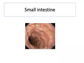
Regional differences of small intestine • Duodenum: Its submucosa has Brunner’s glands. It is invested by serosa or adventitia • Jejunum: has neither Brunner’s glands nor Peyer’s patches. It is invested by serosa. • Ileum: Its lamina propria, opposite the attachment of the mesentery, has lymphoid nodules (Peyer's patches) that extend to the submucosa. It is invested by serosa.