
Histopathological Analysis of HNGC-2 Xenografts in Nude Mice
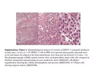
This study examines the histopathological characteristics of HNGC-2 xenografts developed in nude mice. Intracranial injection of as few as 1×10^3 HNGC-2 cells in PBS led to tumor formation in the brain parenchyma, dissected between days 8 and 14. Photomicrographs reveal hypercellularity, a fibrillary background, and palisading necrosis (A). Higher magnification highlights cellular pleomorphism and necrosis (B), while atypical mitosis in tumor cells is observed at high magnification (C).

Histopathological Analysis of HNGC-2 Xenografts in Nude Mice
E N D
Presentation Transcript
Supplementary Figure 1. Histopathological analysis of sections of HNGC-2 xenografts produced in nude mice. As few as 1×10³HNGC-2 cells in PBS were injected intracranially into nude mice (n=4) and tumors developed in the brain parenchyma were dissected out between 8-14 days. (A) The photomicrograph of H&E stained sections show an hypercellular tumor with cells arranged on fibrillary background and palisading necrosis marked by arrow (H&EX40). (B) Higher magnification showing the cellular pleomorphism and necrosis (H&EX100). (C) Tumor cells showing atypical mitosis(H&EX400).