Download
1 / 85
1.03k likes | 3.08k Vues
Neurosensory: Altered Cerebral Function and Increased intracranial pressure (IICP). Updated Fall 2011 by John Nation, RN, MSN From the notes of Charlene Morris, RN, MSN & Marnie Quick, RN, MSN, CNRN. Overview of Today’s Lecture. Discuss altered cerebral function Anatomy and physiology

E N D
Neurosensory:Altered Cerebral Function and Increased intracranial pressure (IICP) Updated Fall 2011 by John Nation, RN, MSN From the notes of Charlene Morris, RN, MSN & Marnie Quick, RN, MSN, CNRN
Overview of Today’s Lecture • Discuss altered cerebral function • Anatomy and physiology • Definition of common terms • Neurological assessment techniques • Increased intracranial pressure • Anatomy and physiology • Clinical manifestations • Interventions • Nursing concerns
Flow of CSF: Produced by filtration of the blood by the choroid plexus of each ventricle flows inferiorly through the lateral ventricles, intraventricular foramen, third ventricle, cerebral aqueduct, fourth ventricle and subarachnoid space and to the blood.
Altered Cerebral Function:Arousal/cognition (LOC) Patho/assessment • Reticular Activating System (RAS) – Reticular Formation - meshwork of gray cell within brainstem extending to the thalamus. • Controls wakefulness, arousal and alertness. • Cerebral cortex outer layer of gray cell bodies of brain. Controls cognition, thought process.
Altered Cerebral Function:What is Consciousness? • Consciousness (Merriam- Webster): waking life (as that to which one returns after sleep, trance, or fever) in which one's normal mental powers are present “the ether wore off and the patient regained consciousness” • Dynamic state • Continuum from awareness of self and environment to unawareness • Consciousness to deep coma • Coma- prolonged unconsciousness
Causes of Changes in LOC • Alcohol intoxication • Drug intoxication (particularly opiates, narcotics, sedatives, and anti-anxiety or seizure medications) • Arrhythmia • Brain disorders • Central nervous system diseases • Lack of oxygen (hypoxia) • Abnormal blood sugars (diabetic coma) • Electrolyte or mineral imbalance • Exposure to heavy metals or hydrocarbons • Extreme fatigue or sleep deprivation • Ketoacidosis • Head trauma • Heart failure • Hypoglycemia (low blood sugar) • Increased carbon dioxide levels (hypercarbia) often seen in emphysema • Infection • Low blood pressure (hypotension) • Metabolic disorders • Thyroid or adrenal gland disorders • Seizures such as those related to epilepsy • Shock • Stroke Source: National Institute of Health
Causes of Coma: Coma can be caused by: • Traumatic brain injuries. Brain injuries that result from traffic collisions or acts of violence are the most common cause of comas. • Stroke. Acute loss of blood flow to the brain followed by swelling or no blood flow to a major part of the brainstem can result in a coma. • Diabetes. Blood sugar levels that get too high (hyperglycemia) and stay too high or get too low (hypoglycemia) and stay too low can cause coma. Source: Mayo Clinic: http://www.mayoclinic.com/health/coma/DS00724/DSECTION=causes
Causes of Coma (Cont’d): • Lack of oxygen. People who have escaped drowning or been resuscitated after a heart attack may not awaken due to lack of blood flow and oxygen to the brain. • Infections. Encephalitis and meningitis are infections that cause inflammation of the brain, spinal cord or the tissues that surround the brain. Severe cases of either encephalitis or meningitis can result in a coma. • Toxins. Exposure to toxins, such as carbon monoxide or drug overdoses, can cause brain damage and coma. Source: Mayo Clinic: http://www.mayoclinic.com/health/coma/DS00724/DSECTION=causes
Altered Levels of Consciousness:Definitions of Terms • Lethargy - a slight reduction in alertness,less aware of what is happening around them and think more slowly. • Obtundation - a moderate reduction in alertness or clouding of consciousness. • Stupor - an excessively long or deep sleeplike state. Arousal is brief by vigorous stimulation, such as repeated shaking, loud calling, pinching. • Coma - is a state of complete unresponsiveness,cannot be aroused, in a deep coma lacks avoidance of pain. • Some reflexes may be present.
Altered Cerebral Function:Assessment of arousal/cognition (LOC) Is the patient alert? • Assess to person/place/time/event (A&O x 4) • Respond to verbal stimuli? • Respond to painful stimuli? • Purpose: shows the brain receives the impulse, interprets it, and responds • Types of painful stimuli: • Trapezius pinch- grasp at least two inches of trapezius muscle. Squeeze and twist. • Supraorbital pressure- carefully applied upward pressure on the ridge along the upper portion of the bony orbital structure • Pressure on finger nails • Sternal rub- not considered appropriate • Is the patient unresponsive? A-----V-----P-----U!
Descending Response to pain stimuli • Pushes your hand away • Pulls away from pain site • General movement • Flexion • Extension • No response
Decorticate posturing- abnormal flexion Decerebrate posturing- abnormal extension
Glasgow Coma Scale A score of 13 to 14 indicates mild deficit. A score between 9 and 12 points to moderate deficit, and a score of 8 or less indicates severe coma.
Assessment of Vital Signs • Temperature - hypothalamus pressure can lead to alterations in body temperature • Cushing’s triad – caused by edema & increased intracranial pressure 1) Increased systolic BP 2) Decreased pulse rate 3) Irregular respirations
Assessment of arousal/cognition - Respiratory • Respiratory- changes occur as brainstem is being compressed • Yawning & sighing • Cheyne-Stokes – crescendo-decrescendo with apnea • Central Neurogenic hyperventilation • Apneustic breathing – Pauses in inspiration and expiration • Cluster breathing – irregular deep to shallow with apnea • Ataxic respirations - grossly irregular
Assessment of arousal/cognition Pupillary light reflex Occipital lobe Brain stem Sensory: CN 2 - Optic Motor: CN 3 - Occulomotor • Note pupil size; darken room; shine light in and note reaction and size
Assessment of arousal/cognition Pupillary light reflex • PERRLA- “Pupils equal, round, reactive to light and accommodation” Anisocoria: The two pupils are not of equal size. Light-near dissociation, refers to a condition where the light reflex is absent or abnormal but the near response is intact. There is no clinical condition in which the light reflex is present and the near response is absent. Amaurotic: blind eye still has consensual response
Assessment Arosual/cognition EOM’S& Brain stem function • Eye movement- CN 3,4,6 • In Deep COMA- test EOM’s by Oculocephalic reflex • Doll’s eyes- Sensory- CN 8; Motor- CN 3,4,6 • Good Dolls eyes: eyes move in opposite direction of head movement – intact brain stem at Pons & nerves • Bad/negative Dolls eyes: eyes do not move head turned How tested with spinal cord injury?
Assessment arousal/cognition Additional Motor Assessment • Ability to move, strength, and symmetry • Grips, arm strength, & drift • Planter flexion, dorsiflexion, & leg strength • Coordination • Finger to nose, heel up and down shin • Planter Reflex- Babinski testing • Meningeal signs- Brudzinski, nuchal rigidity
Planter Reflex and Babinski testing Babinski's reflex – present when the great toe flexes toward the top of the foot and the other toes fan out after the sole of the foot has been firmly stroked. • Postitive response indicates damage to nerve paths connecting the spinal cord and the brain (corticospinal tract) • Abnormal after the age of 2.
Meningeal signs- Brudzinski, nuchal rigidity One of the physically demonstrable symptoms of meningitisis Brudzinski's sign. Severe neck stiffness causes a patient's hips and knees to flex when the neck is flexed.
Meningitis signs- Kernig’s sign Kernig's sign. Severe stiffness of the hamstrings causes an inability to straighten the leg when the hip is flexed to 90 degrees.
Neuro assessment - Sensation • Dull vs. sharp – use broken tongue depressor or cotton tip applicator • Include face, hands, arms, abdomen, feet, and legs
Neuro Assessment Videos • Lewis DVD
Altered cerebral function Nursing assessment for Cerebral Dysfunction • Terms used to describe LOC • Description more important than term • Health history- drugs/head injury/metabolic • Physical exam- modify as individual cooperation • Neuro Vital Signs • LOC, V/S, Pupils, Strength/Movement, Sensation • Glasgow coma scale • NIH Stroke Scale – want low score • NIH Stroke Scale pdf
Common manifestations/Complications Coma states and brain death • Irreversible coma- persistent vegetative state • Does not have functioning cerebral cortex • Caused by anoxia or severe brain injury • Sleep-wake cycles; chew/swallow/cough, no tracking • Locked-in Syndrome (not true coma) • Functioning RAS & cortex; pons level interference • Aware, communicate with eyes • Brain death • Loss of all brain function- flat EEG, no blood flow
Prognosis of individual with altered cerebral functioning • Outcome varies according to underlying cause and pathologic process • The longer the individual unconscious, the longer has absent Doll’s eyes; the poorer the cognitive recovery • Residual mental problems typically outweigh the physical
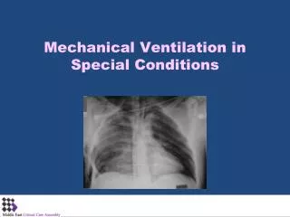
Altered Cerebral FunctionTherapeutic Interventions • Diagnostic tests- to R/O & identify cause of altered cerebral function • CT, MRI, EEG, blood work • Medications- vary according to problem • Overdose; fluid/electrolyte replacement; antibiotics • Surgery- (Ex. tumors, intracranial bleeds) • Other- airway/vent; treat IICP; enteral feeding
Altered Cerebral Functioning:Pertinent Nursing problems Identify the priorities: • Impaired physical mobility • Risk for aspiration • Ineffective coping- Family • Ineffective tissue perfusion (cerebral) • Risk for impaired skin integrity • Ineffective airway • Risk for imbalanced nurtition • Alteration in breathing pattern • Home care
Increased Intracranial Pressure- Overview • Normal ICP Control • Autoregulation • Causes of Increased ICP • Clinical Manifestations • Diagnostic Studies • Monitoring Increased ICP • Treatment
ICP • Skull is a closed box with three essential components: blood 12%, brain tissue 78%, and cerebrospinal fluid (CSF) 10% • Normally, arterial pressure, venous pressure, intraabdominal and intrathoracic pressure, posture, temperature, and blood gases keep ICP relatively constant
Monro-Kellie hypothesis • Brain tissue, blood, and CSF are mostly constant in volume • If the volume of one component increases, another component will be displaced • In total, the intracranial pressure will not change while compensation is possible • (ex. Change in CSF production or absorption, vasoconstriction or dilation, compression or distention of brain tissue) • The ability to accommodate change is limited
Measuring Increased ICP • Normal ICP is 0 to 15 mm Hg • Usually treated once above 20 mm Hg
Cerebral Blood Flow (CBF) • Cerebral Blood Flow- amount of blood in mls passing through 100g of brain tissue in 1 minute • Global CBF is 50ml/min • Normal blood flow 25ml/min in white matter • Normal blood flow 75 ml/min in gray matter • Brain requires constant supply of oxygen and glucose • Brain uses 20% of oxygen and 25% of glucose
Autoregulation • Autoregulation- the automatic adjustment in diameter of cerebral blood vessels to maintain constant blood flow despite changes in blood pressure • Autoregulation is not effective with a MAP less than 50 mm Hg • Autoregulation is not effective with a MAP greater than 150 mm Hg