Download
1 / 66
660 likes | 685 Vues
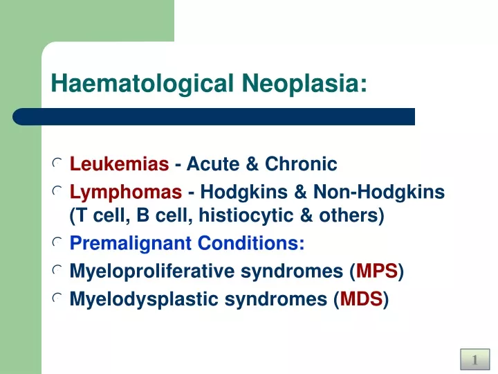
Haematological Neoplasia:. Leukemias - Acute & Chronic Lymphomas - Hodgkins & Non-Hodgkins (T cell, B cell, histiocytic & others) Premalignant Conditions: Myeloproliferative syndromes ( MPS ) Myelodysplastic syndromes ( MDS ). Lymphomas. Introduction:. Neoplastic lymphoid proliferation

E N D
Haematological Neoplasia: • Leukemias - Acute & Chronic • Lymphomas - Hodgkins & Non-Hodgkins (T cell, B cell, histiocytic & others) • Premalignant Conditions: • Myeloproliferative syndromes (MPS) • Myelodysplastic syndromes (MDS)
Introduction: • Neoplastic lymphoid proliferation • Two types – Hodgkins & Non-Hodgkins. • Fever, lymphadenopathy, • Immunodeficiency / autoimmune syndrome • Viral, genetic, unknown etiology. • Lack of programmed cell death - Apoptosis
Lymphoma’sWhere They Begin • Lymphomas are a cancer of the lymphatic system • Lymphatic vessels • Lymph nodes (underarms, groin, neck, spleen, tonsils and bone marrow)
Lymphoma’sWhere They Begin • The Lymphatic system is our bodies main fight against infection • Lymphocytes (B-cell and T-cell) • Carried through our lymphatic system and help our bodies fight infection • Lymphocytes are carried through the lymph vessels as well as the blood stream, so cancer can start in nodes and spread anywhere throughout the body.
Lymphoma Row of enlarged lymph nodes
Hodgkin’s Disease • Is a malignant lymphoma characterized by the presence of atypical, multinucleated giant cell (Reed-Sternberg cell) • The disease is slightly more common in men • Most patients have asymptomatic lymphadenopathy at the time of diagnosis • The initial site of nodal involvement is: • Cervical (65-80%) • Axillary (10-15%) • Inguinal (6-12%)
Causes/Risk Factors • The exact cause of Hodgkin’s disease is unknown. • Also, a virus called Epstein-Barr may be involved with an increased risk of Hodgkin’s. • Also, Agent Orange, used during the Vietnam War is linked with the development of Hodgkin’s. • Siblings of a Hodgkin’s victim are more at risk for developing the disease. • It is more common for men to have the disease. • Hodgkin’s occurs most often in those between 15 and 34 or over the age of 55.
Classification of Hodgkin’s Disease • Treatment depends on • The anatomic distribution of the disease and the presence or absence of specific symptoms • The stage of the disease • The histopathologic subtype
Classification of Hodgkin’s Disease • The Rye classification • Based on four histopathologic subtypes • Lymphocytes predominance • Nodular sclerosis • Mixed cellularity • Lymphocytes depletion
Classification of Hodgkin’s Disease • Histopathologic diagnosis • Is made by lymph node biopsy • Nodes from the lower cervical or axillary areas provide the best tissue for evaluation • Ann Arbor staging classification • A classification based on anatomic distribution of the disease • Stage I disease – Stage IV disease
Splenic Involvement • The probability of splenic involvement increases with increasing spleen size • The absence of splenomegaly does not exclude splenic involvement • Upon gross examination of the spleen a grayish white nodule ranging from several millimeters to several centimeters is apparent with Hodgkin’s disease • Liver involvement with Hodgkin’s disease rarely occurs in the absence of splenic disease
Classifications • Classic Hodgkin’s divisions • Nodular Sclerosing • Mixed Cellularity • Lymphocyte rich • Lymphocyte depleted
Nodular Sclerosing • >60% of cases • Connective tissue • RS cells • Lacunar cells • other cells
Mixed Cellularity • 15 – 30% cases • Mixed cells • Lymphocytes • Histocytes • Eosinophils • Plasma cells • RS cells • Variants • +Necrosis
Lymphocyte rich • <5% cases • Large numbers of Lymphocytes • May suppress other cells • RS cells • Not to be confused with LP Hodgkin’s
Lymphocyte depleted • <1% cases • Lymphocytes rare • RS cells predominate
Symptoms & Presentation • Swollen, painful lymph nodes • Fatigue, etc. • Generalized iching • Appetite, weight loss • Neck %/or back pain • Hair loss • Night sweats
Hodgkin’s Disease • Symptoms such as fever, night sweats, weight loss and pruritus are indicative of widespread involvement and are unfavorable prognostic signs • A typical fever pattern is a high-temperature alternating for a few days with an afebrile period • Many patients have a mild normochromic, normocytic anemia • 1/3 have a leukocytosis due to a neutrophil increase • Eosinophilia is frequently present
Prognosis • Staging • Nodular or Diffuse • Anatomical Staging • Stage I- one region • Stage II- two regions, same side diaphragm • Stage III- two regions, both sides of diaph • Stage IV- spread outside of lymph system • I & II- >10 years • III & IV - < 6 years
Current Treatment of Hodgkin’s Disease • Integrates radiation therapy and combination chemotherapy to achieve the maximum potential for cure • Untreated Hodgkin’s has a five-year survival rate of 5% • The Stanford experience-patients at all stages have survival rate of 86% • Patients in stage IV have a generally poor prognosis
NON- Hodgkin’s Lymphoma • Absence of Reed-Sternberg Cells • May result from damage to DNA that controls growth of cells in immune system • Increased incidence in immunodeficiency
Associated with Non-H lymphoma • SLE • Celiac Disease • AIDS • Organ transplant patients • Rheumatoid Arthritis
Causes and Risk Factors • The Exact causes are still unknown • Higher risk for individuals who: • Exposed to chemicals such as pesticides or solvents • Infected w/ Epstein-Barr Virus • Family history of NHL (although no hereditary pattern has been established) • Infected w/ Human Immunodeficiency Virus (HIV)
Classifications • Based on cell type and location(s) • Non symptomatic -> • Aggressive • Worst prognosis < 1 year
Non-Hodgkin’s Lymphoma (NHL) • A diverse group of primary malignancies of lymphoreticular tissue • The clinical course and natural history is more variable than Hodgkin’s disease • The pattern of spread is irregular and more patients have leukemic features • Current histologic classifications utilize electron microscopic morphology, histochemical studies and selected cell surface antigens. • For our purposes NHL is classified according to nodular (favorable) and diffuse (unfavorable) types
NHL • In contrast to Hodgkin’s disease, about two-thirds of patients initially have asymptomatic lymphadenopathy • In addition to peripheral and mediastinal lymphadenopathy NHL is commonly found initially as an abdominal mass or as hepatic or splenic enlargement • Fever, weight loss and night sweats are frequently present • The median age at diagnosis is 50 • No sex preference is noted
NHL • Patients below 35 or over 65 are more likely to have diffuse histology • As with Hodgkin’s, chemotherapy and / or radiation therapy are the primary forms of treatment • Staging laparotomy is seldom required • Significant therapeutic benefit can be achieved by splenectomy in 80-90 % of patients with advanced lymphomas (including Hodgkin’s disease)
Classification • Usually classified by how the cells look under a microscope and how quickly they grow and spread • Aggressive lymphomas (high-grade lymphomas) • Indolent Lymphomas (low-grade lymphomas)
Classification of Malignant Lymphomas • Low Grade: small lymphoid cells, nodular growth • Intermediate Grade: large cells, follicular and diffuse patterns • High Grade: immunoblastic, lymphoblastic, Burkitt’s disease • T-cell lymphomas: peripheral, cutaneous
Non-Hodgkin’s LymphomaStaging • Stage is the term used to describe the extent of tumor that has spread through the body( I and II are localized where as III and IV are advanced. • Each stage is then divided into categories A, B, and E • A: No systemic symptoms • B: Systemic Symptoms such as fever, night sweats and weight loss • E: Spreading of disease from lymph node to another organ
Non-Hodgkin’s Lymphoma • Two main types of Non-Hodgkin’s Lymphoma: • B-Cell and T-Cell Lymphomas • B-Cell lymphomas (80%) • T-Cell lymphomas (15%)
NHL – Classification: • According to cell type • T cell, B cell, Histiocytic & Misc. NHL • According to Clinical grade • Low grade, Intermediate & High grade NHL. • Histopathological • Diffuse/Follicular NHL, • Small, Intermediate & Large cell NHL Ex: Lennert’s lymphoma is a low grade Tcell NHL. Burkitt’s lymphoma is a high grade B cell NHL
Kiel Classification of NHL • B Cell NHL: • Low Grade: lymphocytic, plasmacytic, centrocytic, mixed centrocytic centroblastic. • High Grade: Centroblastic, Immunoblastic, Burkitts, lymphoblastic. • T Cell NHL: • Low Grade: lymphocytic, mycosis, Lennerts • High Grade: immunoblastic, lymphoblastic etc. • Rare types:
NCI – Working Formulation • Low-grade NHL: • Small lymphocytic • Follicular small cleaved • Intermediate-grade NHL: • Follicular large cell • Diffuse small cleaved • High-grade NHL: • Immunoblastic • Lymphoblastic • Miscellaneous: Histiocytic, Mycosis etc.
NHL- Histologic types Diffuse - & - Follicular
NHL- Histologic types Small – Intermed. – Large