Download

1 / 18
180 likes | 200 Vues
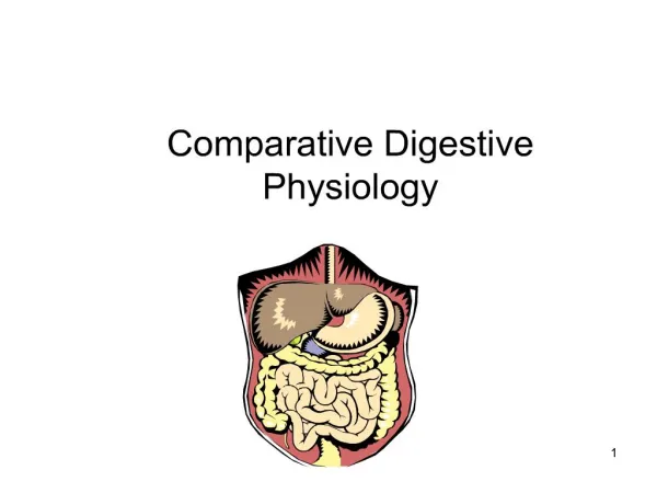
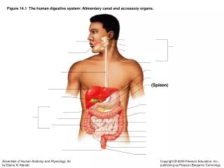
OTHER AMEBAE INHABITING THE ALIMENTARY CANAL Most of these amoebae are commensal organisms that can parasitize the human gastrointestinal tract. Entamoeba hartmanni :

E N D
OTHER AMEBAE INHABITING THE ALIMENTARY CANAL Most of these amoebae are commensal organisms that can parasitize the human gastrointestinal tract. Entamoeba hartmanni: In all of its life–cycle stage, E.hartmanni resembles E.histolytica except in size, yet there is a slight overlap in the size range. The trophozoites do notingest red blood cells, and their motility is generally less vigorous than that of E.histolytica. As in other amebae, infection is acquired by ingestion of food or watercontaminated with cyst-bearing faeces. Identification is based on examination of smallamebae in unstained or iodine-stained preparations. Usually no treatment is indicated,measures generally effective against faecal-borne infections will control this amoebic infection.
The life cycle stages include; trophozoite, precyst, cyst, metacyst, and metacystic trophozoite. Typically the movements of trophozoites are sluggish, with broad short pseudopodia and little locomotion, but at a focus the living specimen cannot be distinguished from the active trophotozoite of E.histolytica. However, the cysts are remarkably variable in size.Entamoeba coli is transmitted in its viable cystic stage through faecal contamination. Ε.coli as a lumen parasite is non-pathogenic and produces no symptoms. The mature cyst (with more than four nuclei) is the distinctive stage to differentiate E.coli from the pathogenic E.histolytica. Specific treatment is not indicated since this amoeba is non-pathogenic. The presence of E.coli in stool specimen is evidence for fecal contamination. Prevention depends on better personal hygiene and sanitary disposal of human excreta.
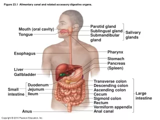
Endolimax nana: Is a lumen dweller in the large intestine, primarily at the cecal level, where it feeds on bacteria. The life cycle is similar to E.histolytica. Motility is typically sluggish (slug-like) with blunt hyaline pseudopodia, Projects shortly. Human infection results from ingestion of viable cysts in polluted water or contaminated food. Typical ovoid cysts of E.nana are confirmative. Rounded cysts and living trophozoites are often confused with E.hartmanni and E.histolytica. No treatment is indicated for this nonpathogenic infection. Prevention can be achieved through personal cleanliness and community sanitation.
Iodamoeba buetschlii: - the natural habitat is the lumen of the large intestine, the principal site probably being the caecum. The trophozoite feeds on enteric bacteria; it is a natural parasite of man and lower primates. It is generally regarded as a nonpathogenic lumen parasite. No treatment is ordinarily indicated. Prevention is based on good personal hygiene and sanitation in the community. Entamoeba gingivalis - Only the trophozoite stage presents, and encystation probably does not occur. E.gingivalis is a commensal, living primarily on exudate from the margins of the gums, and thrives best on unhealthy gums. No specific treatment is indicated. However the presence of E.giingivalis suggests a need for better oral hygiene. The infection can be prevented by proper care of the teeth and gums.
Blastocystis hominis- is an inhabitant of the human intestinal tract previously regarded as non-pathogenic yeast. Its pathogenecity remains controversial. The organism is found in stool specimen from asymptomatic people as well as from people with persistent diarrhoea. B.hominis is capable of pseudopodia extension and retraction, and reproduces by binary fission or sporulation. The classic form that is usually seen in the human stool specimen varies tremendously in size, from 6-40μm. There are thin –walled cysts involved in autoinfection, and thick–walled cysts responsible for external transmission via the fecal-oral route.. The organism may be detected in wet mounts or trichome–stained smears of fecal specimens.
2-FLAGELLATES Flagellates are unicellular microorganisms. Their locomotion is by lashing a tail-like appendage called a flagellum or flagella and reproduction is by simple binary fission. There are three groups of flagellates: A- Luminal flagellates Giardia lamblia Dientmoeab fragilis B- Hemoflagellates Trypanosoma species. Leishmania species. C- Genital flagellates Trichomonas vaginalis A- Luminal flagellates
Giardia lamblia The life cycle consists of two stages, the trophozoite and cyst. The trophozoite is 9-12 μm long and 5-15μm wide anteriorly. It is bilaterally symmetrical, pear-shaped with two nuclei (large central karyosome),four pairs of flagella, two axonemes, and a suction disc with which it attaches to the intestinal wall.The oval cyst is 8-12μm long and7-10μm wide, thick-walled with four nucleus and several internal fibera.Each cyst gives rise to two trophozoites during excystation in the intestinal tract. Transmission is by ingestion of the infective cyst.
Pathogenesis Infection with G.lamblia is initiated by ingestion of cysts. Gastric acid stimulates excystation, with the release of trophozoites in duodenum and jejunum. The trophozoites can attach to the intestinal villi by the ventral sucking discs without penetration of the mucosa lining, but they only feed on the mucous secretions. In symptomatic patients, however, mucosa-lining irritation may cause increased mucous secretion and dehydration. Metastatic spread of disease beyond the GIT is very rare.
Clinical features Clinical disease: Giardiasis Symptomatic giardiasis ranges from mild diarrhea to severe malabsorption syndrome. Usually, the onset of the disease is sudden and consists of foul smelling, watery diarrhea, abdominal cramps, flatulence, and streatorrhoea. Blood & pus are rarely present in stool specimens, a feature consistent with the absence of tissue destruction.
Laboratory diagnosis Examination of diarrhoeal stool- trophozoite or cyst, or both may be recovered in wet preparation. In examinations of formed stool (e.g. in asymptomatic carriers) only cysts are seen.Giardia species may occur in “showers”, i.e. many organisms may be present in the stool on a given day and few or none may be detected the next day. Therefore one stool specimen per day for 3 days is important. If microscopic examination of the stool is negative in a patient in whom giardiasis is highly suspected duodenal aspiration, string test (entero-test), or biopsy of the upper small intestine can be examined. In addition to conventional microscopy, several immunologic tests can be implemented for the detection of parasitic antigens. Prevention - Asymptomatic reservoirs of infection should be identified & treated. - Avoidance of contaminated food and water. - Drinking water from lakes and streams should be boiled, filtered and/or iodine treated. - Proper waste disposal and use of latrine.
Trichomonas vaginalis Important features- it is a pear-shaped organism with a central nucleus and four anterior flagella; and undulating membrane extends about two-thirds of its length. It exists only as a trophozoite form, and measured 7-23μm long & 5-15μm wide. Transmission is by sexual intercourse. Pathogenesis The trophozoite is found in the urethra & vagina of women and the urethra & prostate gland of men. After introduction by sexual intercourse, proliferation begins which results in inflammation & large numbers of trophozoites in the tissues and the secretions. The onset of symptoms such as vaginal or vulval pruritus and discharge is often sudden and occurs during or after menstruation as a result of the increased vaginal acidity. The vaginal secretions are liquors, greenish or yellowish, sometimes frothy, and foul smelling. Infection in the male may be latent, with no symptoms, or may be present as self limited, persistent, or recurring urethritis.
Clinical features Clinical disease - trichomoniasis. Most infected women at the acute stage are asymptomatic or have a scanty, watery vaginal discharge. In symptomatic cases vaginitis occurs with more extensive inflammation, along with erosion of epithelial lining, and painful urination, and results in symptomatic vaginal discharge, vulvitis and dysuria.
Laboratory diagnosis • In females, T.vaginalis may be found in urine sediment, wet preparations of vaginal secretions or vaginal scrapings. • In males it may be found in urine, wet preparations of prostatic secretions or following massage of the prostate gland. • Contamination of the specimen with feces may confuse T.vaginalis with T.hominis. Prevention - Both male & female sex partners must be treated to avoid reinfection - Good personal hygiene, avoidance of shared toilet articles & clothing. - Safe sexual practice.
Dientamoeba fragilis Dientamoeba fragilis was initially classified as an amoeba; however, the internal structures of the trophoziote are typical of a flagellate. No cyst stage has been described. The life cycle are simple . It has worldwide distribution. The transmission is postulated, via helminthes egg such as those of Ascaris and Enterobius species. Transmission by fecal- oral routes does occur.
Most infection with D. fragilis is asymptomatic, with colonization of the cecum and upper colon. However, some patients may develop symptomatic disease, consisting of abdominal discomfort, flatulence, intermittent diarrhea, anorexia, and weight loss. The reservoir for this flagellate and lifecycle are unknown. Thus, specific recommendation for prevention is difficult. However, infection can be avoided by maintenance of adequate sanitary conditions