Download
1 / 76
780 likes | 1.09k Vues
Stasis Dermatitis and Leg Ulcers. Medical Student Core Curriculum in Dermatology. Last updated June 8, 2011. Module Instructions.

E N D
Stasis Dermatitis and Leg Ulcers Medical Student Core Curriculum in Dermatology Last updated June 8, 2011
Module Instructions • The following module contains a number of blue, underlined terms which are hyperlinked to the dermatology glossary, an illustrated interactive guide to clinical dermatology and dermatopathology. • We encourage the learner to read all the hyperlinked information.
Goals and Objectives • The purpose of this module is to help medical students develop a clinical approach to the evaluation and initial management of patients presenting with stasis dermatitis and leg ulcers. • By completing this module, the learner will be able to: • Recognize the clinical presentation of stasis dermatitis • List treatment and preventative measures for stasis dermatitis • List the most frequent causes of leg ulcers and describe their presentations • Describe proper wound care and treatment for leg ulcers • Discuss when to refer a patient with leg ulcers to a specialist
Case One Mrs. Lillian Paulsen
Case One: History • HPI: Mrs. Paulsen is a 74-year-old woman who presents to the dermatology clinic with leg discoloration for the past three months. The “rash” does not hurt, but occasionally itches. She has not tried any treatment. • PMH: diabetes (last hemoglobin A1c was 6.7), hypertension, obesity. No history of atopic dermatitis. • Medications: ACE-inhibitor, thiazide diuretic, sulfonylurea • Allergies: none • Family history: noncontributory • Social history: lives with her husband in a nearby town • Health-related behaviors: no tobacco, drug use, or alcohol • ROS: no leg pain when walking or at rest
Case One, Question 1 How would you describe her skin exam?
Case One, Question 1 • Erythematous brown hyperpigmented plaque with fine fissuring and scale located above the medial malleolus on the left lower leg • Right leg with varicosities • Notice the asymmetry? Palpation of the left leg reveals firm skin suggestive of fibrosis
Case One, Question 2 • What is the most likely diagnosis? • Atopic dermatitis • Cellulitis • Erysipelas • Stasis dermatitis • Tinea corporis
Case One, Question 2 Answer: d • What is the most likely diagnosis? • Atopic dermatitis(adults with AD have a history of childhood AD and a different distribution of skin involvement) • Cellulitis(cellulitis occurs more acutely, presents with fever and pain, more erythema, well-demarcated and without pruritus or scale) • Erysipelas(a form of cellulitis caused by acute beta-hemolytic group A streptococcal infection of the skin) • Stasis dermatitis • Tinea corporis(would expect sharply marginated, erythematous annular patches with central clearing)
Diagnosis: Stasis Dermatitis • Stasis dermatitis typically presents with erythema, scale, pruritus (itching), erosions, exudate, and crust • Usually located on the lower third of the legs, superior to the medial malleolus • Can occur bilaterally or unilaterally • Lichenification may develop • Edema is often present, as well as varicose veins and hemosiderin deposits (pinpoint yellow-brown macules)
Venous Insufficiency • Stasis dermatitis is a cutaneous marker of venous insufficiency. • Normally, venous blood returns from the superficial venous system via perforating veins into the deep venous system. • Venous stasis occurs when the valves in the deep or perforating veins become incompetent, causing reflux into the superficial system (venous hypertension).
Venous Insufficiency • Risk factors for venous insufficiency: • Heredity • Age (older) • Female • Pregnancy • Chronic venous disease is extremely common and is associated with a reduced quality of life secondary to pain, decreased physical function, and mobility • Obesity • Prolonged standing • Greater height
Venous Insufficiency • Early signs of venous insufficiency: • Tenderness • Edema • Hyperpigmentation • Late signs: • Lipodermatosclerosis(subcutaneous fat is replaced by fibrosis that eventually impedes venous and lymphatic flow leading to edema above the fibrosis) • Venous ulcers • Scars that appear porcelain white and atrophic • Telangiectasias • Varicose veins
Lipodermatosclerosis • Stasis dermatitis can lead to fat necrosis with the end stage being permanent sclerosis (lipodermatosclerosis) with “inverted champagne bottle” legs as seen here • Patients with lipodermatosclerosis may also have acute inflammatory episodes that present with pain and erythema (these episodes can be mistaken for cellulitis)
Elephantiasis Verrucosa Nostra • Inflammation of the draining lymphatics (as occurs with cellulitis) results in damage to those vessels resulting in lymphatic insufficiency • The overlying skin becomes pebbly, hyperkeratotic, and rough • Ulceration in this setting (with lymphatic and venous insufficiency) is significantly harder to treat and heal
Case One, Question 2 • Which of the following are complications of venous insufficiency? • Cellulitis • Contact dermatitis • Recurrent ulceration • Venous thrombosis • All of the above
Case One, Question 2 Answer: e • Which of the following are complications of venous insufficiency? • Cellulitis • Contact dermatitis • Recurrent ulceration • Venous thrombosis • All of the above
Complications of Venous Insufficiency • Recurrent ulcers • Cellulitis (open wound provides a portal of entry for bacteria) • Contact dermatitis (from topical agents applied to stasis dermatitis or ulceration) • Venous thrombosis
Leg Ulcers and Contact Dermatitis • Leg ulcers are subject to sensitization to products used to treat wound healing, leading to contact dermatitis. • This is due to the intrinsic allergenic properties of many ointments and wound products, the duration of use, and the disrupted skin barrier. • This chronic inflammation and resultant dermatitis lead to poor wound healing and/or recurrence of leg ulcers.
Stasis Dermatitis: Treatment • It is important to treat both the dermatitis and the underlying venous insufficiency • Application of super-high and high potency steroids to area of dermatitis • Elevation (to reduce edema) • Compression therapy with leg wraps • Change wraps weekly, or more often if the lesion is very weepy
Compression Therapy Works PRIOR TO TREATMENT FOLLOWING TREATMENT
Case Two Mr. Patrick Baily
Case Two: History • HPI: Mr. Baily is a 50-year-old man who presents to his primary care provider with pain in his left leg. He developed a “weeping spot” a few weeks ago, which he tried treating with an over-the-counter antibiotic ointment. • PMH: history of a DVT 5 years ago after a transatlantic flight, no longer on anticoagulation, hypertension, type 2 diabetes • Medications: thiazide diuretic, ACE-inhibitor, glyburide, metformin • Allergies: none • Family history: father with type 2 diabetes and hypertension • Social history: lives with wife in an apartment, works in construction • Health-related behaviors: smokes 1 cigarette/day • ROS: as above
Case Two, Question 1 • How would you describe Mr. Baily’s skin exam?
Case Two, Question 1 • Irregularly shaped ulcer located on the medial aspect of the left ankle, erythematous border, exudative • Without undermining (unable to probe under the edges) • Pedal pulses are present, 1+
Case Two, Question 2 • Given the history and exam, what type of ulcer is on Mr. Baily’s left leg? • Arterial • Diabetic • Pressure • Venous
Case Two, Question 2 Answer: d • Given the history and exam, what type of ulcer is on Mr. Baily’s left leg? • Arterial • Diabetic • Pressure • Venous
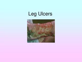
Venous Insufficiency Ulcers • Active or healed venous leg ulcers occur in ~ 1% of the general population • They typically appear as tender, shallow, irregular ulcers with a fibrinous base that are always located below the knee • Usually located on the medial ankle or along the line of the long or short saphenous veins • Accompanied with leg edema, hemosiderin pigmentation, +/- dermatitis of the leg • Patients may experience symptoms of aching or pain. Discomfort may be relieved by elevation.
Leg Ulcers • Causes of chronic leg ulcers include: • Venous insufficiency 45-60% • Arterial insufficiency 10-20% • Combination of venous and arterial 10-15% • Diabetic 15-25% • Malignancy, vasculitis, collagen-vascular diseases, and dermal manifestations of systemic disease may present as ulcers on the lower extremity • Smoking and obesity increase the risk for ulcer development and persistence (independent of the underlying cause)
Case Two, Question 3 • Which of the following is the most appropriate next step in evaluating Mr. Baily? • Measure the blood pressure in the left arm and left ankle • Obtain a skin biopsy • Treat the ulcer with topical antibiotics • Use electrocautery to stop the weeping
Case Two, Question 3 Answer: a • Which of the following is the most appropriate next step in evaluating Mr. Baily? • Measure the blood pressure in the left arm and left ankle (Mr. Baily’s DP pulse was weak suggesting possible co-existent peripheral arterial disease) • Obtain a skin biopsy (not necessary unless the diagnosis is unclear or the ulcer does not respond to treatment) • Treat the ulcer with topical antibiotics (no, in fact topical antibiotic ointments may lead to a contact dermatitis) • Use electrocautery to stop the weeping (trauma may worsen the wound instead of improve it)
Ankle/Brachial Index (ABI) • Measure the ABI to exclude arterial occlusive disease • Compression therapy (used to treat venous insufficiency) is contraindicated in patients with significant arterial disease • The ABI is the ratio of systolic blood pressure in the ankle to the systolic blood pressure in the brachial artery • Normal: ≥ 0.8 • < 0.8 = indication of peripheral arterial disease
Ankle/Brachial Index (ABI) • The ABI is reliable except in diabetes (may be falsely high) • An ABI should be performed in all patients with weak peripheral pulses, risk factors for arterial occlusive disease (e.g. smoking, diabetes, hyperlipidemia), and when ulcers are in locations not consistent with venous ulcers
Venous Ulcers: Evaluation • In addition to assessment of the ulcer, the physical exam of patients with leg ulcers should include the evaluation of peripheral pulses, capillary refill time, peripheral neuropathy, and deep tendon reflexes • Diagnosis of venous leg ulcers can be made clinically, however, non-invasive vascular studies such as venous duplex ultrasound and venous rheography can help document the presence and etiology of venous insufficiency • Findings may warrant surgical intervention with endoscopic venous laser ablation, which may prevent further complication • Surgical intervention tends to be more helpful when the venous disease is limited
Venous Ulcers: Treatment • Address the underlying cause (venous insufficiency) as well as local wound care: • Leg elevation • Keep the wound moist with a primary dressing • Treat dermatitis with topical steroids • Compression therapy (except with an ABI < 0.8) • Apply external compression (applied over a primary dressing) with a high compression system such as a multilayer bandage or paste-containing bandage (e.g. Unna’s boot, Duke boot) • Treat infection with debridement of necrotic or infected tissues and use systemic antibiotics for infection • Measure the ulcer at each visit to document improvement
Wound Care: The Primary Dressing • Keep the wound moist. A moist wound environment promotes healing compared to air exposure • Choice of dressings is less important than the program of ulcer treatment outlined on the previous slide • Semipermeable dressings that allow oxygen and moisture to pass through (but not water) have made the treatment of leg ulcers easier and more effective
Venous Ulcers: Treatment • Patient education is crucial in successful treatment: • Avoid topical antibiotics in order to prevent sensitization and development of contact dermatitis • Cleanse the wound with saline. Avoid products like betadine and hydrogen peroxide to prevent skin breakdown • Avoid frequent manipulation of the wound. Dressings can be changed as infrequently as once weekly. • Once healed, avoid reaccumulation and development of ulcers with regular use of 20-30mmHg compression stockings • Patients with venous ulcers that do not demonstrate response to treatment (reduction in size) after 6 weeks should be referred to dermatology or a wound care clinic
Case Three Mr. Robert Lund
Case Three: History • HPI: Mr. Lund is a 60-year-old man who presents to his primary care provider with a painful “sore” on his right lateral leg. He reports a history of a “cramping pain” in his calves when walking, but this current pain is more localized to the skin. • PMH: hyperlipidemia, hypertension, angina (stable) • Medications: statin, thiazide diuretic, sublingual nitroglycerin when needed, aspirin • Allergies: NKDA • Family history: father with an MI at age 65, mother with diabetes • Social history: lives with his wife, works in sales, 2 grown children • Health-related behavior: smokes ½ pack of cigarettes/day, one glass of wine nightly, no drug use • ROS: no shortness of breath or recent chest pain
Case Three, Question 1 • How would you describe Mr. Lund’s skin exam?
Case Three, Question 1 • “Punched out” appearing ulcer with sharply demarcated borders • Minimal exudation and surrounding erythema • Dorsalis pedis pulse is absent • ABI is 0.6
Arterial Ulcers • Arterial ulcers are caused by peripheral arterial disease • Occur on the lower leg, usually over sites of pressure and trauma: pretibial, supramalleolar, and at distant points, such as toes and heels • Appear “punched out,” with well-demarcated edges and a pale base • Exudation is minimal • Associated findings of ischemia include loss of hair on feet and lower legs, shiny atrophic skin
Arterial Ulcers • Pulses (dorsalis pedis and posterior tibial) may be diminished or absent • Stasis pigmentation and lipodermatosclerosis are absent (unless patient also has venous disease) • Associated with intermittent claudication and pain • As disease progresses, pain and claudication may occur at rest • Unlike venous ulcers, leg pain often does not diminish when the leg is elevated
Case Three, Question 2 • Which of the following recommendations should take priority? • Encourage him to ambulate • Encourage him to stop smoking • Make sure his blood pressure and hyperlipidemia are under good control • Refer to a vascular surgeon
Case Three, Question 2 Answer: d • Which of the following recommendations should take priority? • Encourage him to ambulate • Encourage him to stop smoking • Make sure his blood pressure and hyperlipidemia are under good control • Refer to a vascular surgeon (although all the answer choices are correct, the main goal of therapy is the re-establishment of adequate arterial supply)
Arterial Ulcers: Treatment • Refer to a vascular surgeon for restoration of arterial blood flow with percutaneous or surgical arterial reconstruction • Patients should stop smoking, optimize control of diabetes, hypertension, and hyperlipidemia • Weight loss and exercise are also helpful • All types of ulcers require proper wound care as outlined above in venous ulcer treatment