
Nervous System
Nervous System. Brain, spinal cord, efferent and afferent neurons Pattern of information flow:. Receptor Afferent path Integration Efferent Path Effect. Central Nervous System (CNS). Main cell types are neurons and glial cells. Typical Arrangement of Neural Connections.

Nervous System
E N D
Presentation Transcript
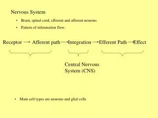
Nervous System • Brain, spinal cord, efferent and afferent neurons • Pattern of information flow: Receptor Afferent path Integration Efferent Path Effect Central Nervous System (CNS) • Main cell types are neurons and glial cells
Typical Arrangement of Neural Connections • Neurons communicate via electrical signaling • They are excitable • Structurally the soma (cell body) has an extensive ER and prominent nucleoli • Long appendages or processes: • Dendrites (receive info) • Axons (deliver info); some are covered by myelin A collection of axons is called a NERVE
Types of glial cells: CNS = oligodendrocytes, astrocytes, microglia, ependymal cells PNS = Schwann cells, satellite cells
Myelin acts as an insulator and inhibits ion movement in the axonal membrane that is surrounds.
Neurons as Excitable Tissue • Excited by altering the resting membrane potential (-90 mV) • Depolarize • Hyperpolarize • Most changes in membrane potential occur through the opening or closing of certain ion channels (they are voltage-gated).
What will happen to the resting membrane potential if the activation gate is opened? How could a cell open this activation gate?
Gates can be chemically opened by neurotransmitters • Gates can be opened via signal transduction mechanisms linked to neurotransmitter binding to receptor • Gates can be opened by stretch, pressure, etc.
Stimulus = anything that can cause the opening or closing of gated channels in a neuronal membrane What happens to the resting membrane potential of the membrane adjacent to the site of Na+ entry? How about here?
The axon hillock (trigger zone) is sensitive to changes in ion concentration and is the site at which an action potential is initiated. An action potential is a self-propagating depolarization of the axonal membrane that initiates at the hillock and runs to the axon terminus without diminishing in strength. What determines whether an action potential will occur or not?
If the graded potential doesn’t change the resting membrane potential enough, the signal from the stimulus will die out and the neuron will not respond with an action potential. The amount of change in membrane potential necessary to generate an action potential is called a threshold stimulus.
If the trigger area of the axon reaches threshold, the influx of Na+ and the generation of the action potential will be repeated over and over again in one direction, at each segment of membrane, down the axon.
What will happen at this area of membrane? What will happen at this area of membrane?
One portion of the membrane has just been depolarized and is relatively insensitive to changes in cation concentration. It is said to be refractory to stimulus. Downstream membrane is at resting potential, and can be influenced by cation influx.
Action potentials cause the release of neurotransmitter from the presynaptic axon terminus
EPSP mV time time mV time IPSP
Neurotransmitter activity is stopped by: diffusion away from the synapse, transport into cells (glial or back into presynaptic neuron), or degradation by specific enzymes.
What will determine whether this postsynaptic neuron will respond?
A B Red neuron is releasing serotonin which causes an IPSP. The neuron is firing at 70 APs/sec Neuron A is releasing dopamine, causing and EPSP. The neuron is firing at 40 APs/sec Neuron B is releasing acetylcholine to create an EPSP. It is firing at 20 APs/sec. What will the outcome be in the postsynaptic cell?
Transmitter Molecule Derived From Site of Synthesis Acetylcholine Choline CNS, parasympathetic nerves Serotonin5-Hydroxytryptamine (5-HT) Tryptophan CNS, chromaffin cells of the gut, enteric cells GABA Glutamate CNS Glutamate CNS Aspartate CNS Glycine spinal cord Histamine Histidine hypothalamus Epinephrine Tyrosine adrenal medulla, some CNS cells Norpinephrine Tyrosine CNS, sympathetic nerves Dopamine Tyrosine CNS Adenosine ATP CNS, periperal nerves


