Download
1 / 12
120 likes | 215 Vues
Pharmacognostical standardization and phytochemical screening of Albizzia lebbeck leaves were assessed. Transverse section of Albizzia lebbeck leaves shows an upper and lower single horizontal layered epidermis followed by spongy parenchyma on both lower and upper surfaces, parenchymatous cells are thin-walled with intercellular space. Bundle sheath is present surrounding vascular bundle consist 8-10 layers of parenchymatous cells. Phytochemical screening of ethyl acetate, ethanolic and water maceration extracts of leaves shows presence of different chemical constituents like alkaloids, tannins, carbohydrates, flavanoids, proteins and Amino acids. Foaming index is less then 100 which indicate presence of saponins in leaves which finally proves by quantitative determination of total saponin 1.4% and total flavanoids 0.6%. Water soluble extractives are more then alcohol soluble extractives show more water soluble constituents in the leaves.

E N D
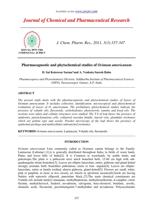
Available on line www.jocpr.com Journal of Chemical and Pharmaceutical Research ____________________________________________________ J. Chem. Pharm. Res., 2010, 2(1): 432-443 ISSN No: 0975-7384 Pharmacognostic standardization and phytochemical screening of albizzia lebbeck Rahul Chulet*1,Lincy Joseph1 , Methew George1, Pankaj Pradhan1 1. Department of Pharmaceutical Sciences Jaipur National University, Jaipur ______________________________________________________________________________ Abstract Pharmacognostical standardization and phytochemical screening of Albizzia lebbeck leaves were assessed. Transverse section of Albizzia lebbeck leaves shows an upper and lower single horizontal layered epidermis followed by spongy parenchyma on both lower and upper surfaces, parenchymatous cells are thin-walled with intercellular space. Bundle sheath is present surrounding vascular bundle consist 8-10 layers of parenchymatous cells. Phytochemical screening of ethyl acetate, ethanolic and water maceration extracts of leaves shows presence of different chemical constituents like alkaloids, tannins, carbohydrates, flavanoids, proteins and Amino acids. Foaming index is less then 100 which indicate presence of saponins in leaves which finally proves by quantitative determination of total saponin 1.4% and total flavanoids 0.6%. Water soluble extractives are more then alcohol soluble extractives show more water soluble constituents in the leaves. Key Words: Mid rib, Phytochemical Screening, Alkaloids, Total Flavanoids ______________________________________________________________________________ Introduction Man has been using herbs and plants products for combating diseases since times immemorial. Indian systems of medicine have a deep root in our culture heritage and cater to the Medicare of large sections of our population. These systems mainly use herbs. If we dwell for a moment on our hoary past, the Rigveda, one of the oldest repositories of human knowledge, mentions the use of 67 plants for therapeutic use, the Yajurveda enlist 81 plants whereas the Atharveda written 432
Rahul Chulet et al J. Chem. Pharm. Res., 2010, 2(1): 432-443 _____________________________________________________________________________ during 1200 BC describes 290 medicinal plants of medicinal value. Charak Samhita written during 990 BC describes 341 medicinal plants. The land mark in Ayurveda was Sushrut Samhita written during 600 BC mentioned 395 medicinal plants. Dhanwantari Nighantu mentions 750 medicinal plants, 450 are mentioned in the Bhavaprakash, 480 in Madanapala Nighantu and 450 in the Kaiyadeva Nighantu. India unquestionably occupies the top position in the use of herbal drugs. It is one of the foremost countries exporting plant drugs and their derivatives. It also excels in home consumption. It is not at all surprising that herbal drugs are so prevalent in India given the great biodiversity and abundance of flora and the variety of geographical condition which allows the most exotic medicinal plants to be grown here. [1] Albizzia lebbeck Benth. (Shirish, Family: Leguminosae) is a deciduous tree with compound leaves, flat oblong fruits, round cream colored seeds, grows wild. The plant is found throughout India, Bangladesh, tropical and subtropical Asia and Africa [2]. Barks are used in toothache and diseases of the gum. Decoction of the leaves and barks are protective against bronchial asthma and other allergic disorders. Barks and seeds are astringent and are given in piles and diarrhea. Ethanolic extract of pods possesses antiprotozoal, hypoglycemic and anticancer properties. The methanolic extract of the pod was investigated for antifertility activity [3, 4]. The plant extract also evaluated in allergic rhinitis [5] and memory and learning of mice [6]. Phytochemical investigations showed that the pod of the Albizzia lebbeck contains 3’, 5 Dihydroxy 4’, 7 dimethoxy flavone, and N- Benzoyl L phenyl alaninol [7]. The beans of the plant contain albigenic acid-a new triterpenoid sapogenin [8]. The plant also contains saponins [9, 10], macrocyclic alkaloids [11], Tannins [12], and flavonols [13]. The decoction of Albizzia lebbeck stem bark was found to be effective against bronchospasm induced by histaminic acid phosphate and shown to exert di-sodium chromoglycate like action on mast cells. [14] Albizzia lebbeck bark extract show the antimicrobial activity. The active constitute of bark extract is anthraquinone glycosides. The main constituent from bark is active against aerobes and mechanism of action is that glycosides cause the leakage of the cytoplasmic constituents.[15] Two new tri-O-glycoside flavonols kaempferol and quercetin were identified from the leaves of Albizzia lebbeck. [16] Albizziahexoside a new hexaglycosylated saponin was isolated from leaves ofAlbizzia lebbeck. [17] Lignins Present in their cell walls have been oxidized with alkaline nitrobenzene. The phenolic acids were present in the range of 8.8-52.7 mg/g of cell wall. [18]The chloroform fraction of methanolic extract of Albizzia lebbeck leaves protected mice against maximal electroshocks. [19]Ethyl ether and alcoholic extracts of leaves of Albizzia lebbeck showed positive reaction against bacterial pathogens i.e. [Staphylococcus aureus] and [Escherichia coli] and fungal pathogen [Candida albicans]. Flavonoid contents like Quercetin and Kaempferol were isolated and identified form the leaves and Flavonoid was found contents (2.40 mg/g). [20]Methanolic extract of leaf and methanolic and water extracts of bark have shown in vitro mast cell stabilizing effect against compound 48/80. [21]The effect of saponin containing n-butanolic fraction (BF) extracted from dried leaves of Albizzia lebbeck on learning and memory was studied in albino mice and significant improvement was observed in the retention ability of the normal and amnesic mice as compared to their respective controls. [22] Pharmacognostic and phytochemical study of Albizzia lebbeck leaves is not available up to present day so this study is helpful to increase attraction towards this plant. 433
Rahul Chulet et al J. Chem. Pharm. Res., 2010, 2(1): 432-443 _____________________________________________________________________________ Materials and Methods Plant collection and identification The leaves of Albizzia Lebbeck were collected from Jaipur in March 2009. A voucher specimen (Voucher No. RUBL 50033) was kept at the Department of Botany, University of Rajasthan after identification of the plant. Extraction of the plant material Plant materials were washed with water and shade dried. The derided leaves were crushed to coarsely powdered by wood-grinder. The powdered material was defatted with petroleum ether (60-80 ºC) and then extracted with ethanol in the ratio of 1:10 of powdered drug and solvent by cold maceration method for 24 hrs. The extract was concentrated for further studies on water bath at 40 ºC. Morphological and microscopical study The pieces of leaves were boiled in a test tube with chloral hydrate for several minutes until completely clarified and then examine them in chloral hydrate solution. After clarification, leaf pieces are divided into two parts with the help of a scalpel or needle, and carefully turn one part. The leaf can be examined from both the dorsal and ventral surfaces [23].Transverse section is obtained by cutting the leaf portion including mid rib with the help of sharp blade and staining is done by safranin to impart a red colour to the lignified tissue. Pharmacognostic standardization Total ash About 3 gm accurately weighed powdered drug was incinerated in a silica dish at a temperature not exceeding 4500C until free from carbon. It was then cooled and weighed. The % w/w of ash with reference to the air-dried drug was calculated. Acid insoluble ash To the crucible containing the total ash was added 25 ml of hydrochloric acid. The crucible was then covered with a watch-glass and the mixture was boiled gently for 5 minutes. The watch- glass was rinsed with 5 ml of hot water and this liquid was added in to the crucible. The insoluble matter was collected on an ash less filter-paper and washed with hot water until the filtrate was neutral. The filter-paper contain the insoluble matter was transferred to the original crucible, dried on a hot-plate and ignite to constant weight. The residue was allowed to cool in a desiccator for 30 minutes and then weighed. Water soluble ash To the crucible containing the total ash, was boiled for 5 min. with 25 ml of water. Insoluble matters were collected on ash less filter-paper, washed with hot water and ignite for 15 min. at temp. Not exceeding 450°C. Weight of insoluble matter was subtracted from the weight of total ash. Alcohol soluble extractive Accurately weighed 4 gm of air-dried powdered drug was macerated with 100 ml of alcohol of the specified strength in a closed flask for 24 h, shaken frequently during first 6 h and allowed to 434
Rahul Chulet et al J. Chem. Pharm. Res., 2010, 2(1): 432-443 _____________________________________________________________________________ stand for 18 h. It was then filtered rapidly, taking precautions against loss of the solvent and 25 ml of the filtrate were evaporated to dryness in a tared flat-bottomed shallow dish and dried at 100 oC to constant weight. The % w/w of alcohol soluble extractive value was calculated with reference to the air-dried drug. Water soluble extractive Accurately weighed 4 gm of air-dried powdered drug was macerated with 100 ml of water in a closed flask for 24 h, shaken frequently during first 6 h and allowed to stand for 18 h. It was then filtered rapidly and 25 ml of the filtrate were evaporated to dryness in a tared flat-bottomed shallow dish and dried at 100°C to constant weight. The % w/w of water soluble extractive value was calculated with reference to the air-dried drug. Loss on drying 10 gm of the drug was weighed accurately in a tared evaporating dish. It was dried at 105°C for 5 hours and weighed. The drying and weighing was continue at 1 hour interval until difference between two successive weighing corresponds to not more than 0.25 percent constant weight was reached. Sulphated ash 1 gm of substances was taken in an accurately weighed crucible, ignited gently at first until a substance is thoroughly charred. Cooled and the residue was moistened with 1 ml of sulphuric acid, heat gently until white fumes was no longer evolved and ignite at 800 0C until black particle were disappeared. Allowed the crucible to cool, few drops of sulphuric acid were added and heat. Ignite as before, allowed to cool and weigh. Operation was repeated until two successive weighing was not differing by more than 0.5 mg. [24] Crude fibre content 2 gm of powder drug was taken in a beaker. Add 50 ml of 10% v/v nitric acid. Heated to boil with constant stirring (till about 30 sec. after boiling start) strain through fine cotton cloth on Buchner funnel. Residue was washed with boiling water, transfer the residue from the cloth to a beaker. 50 ml of 2.5% v/v sodium hydroxide was added and heat to boil. Maintain at boiling point for 30 sec. with constant stirring. Strain and washed with hot water as mentioned earlier. Residue was transfer in crucible and weighs the residue and percentage of crude fiber was determined. Foaming index 1 gm coarse powder was weighed accurately and transferred to a 500 ml conical flask containing 100 ml of water. It was maintained at moderate boiling for 30 minute. It was cool and filtered in to a 100 ml volumetric flask, Volume was diluted by adding sufficient amount of water. The decoction was poured in to 10 stoppered test tubes in successive portion of 1 ml, 2 ml, 3 ml -- up to 10 ml and the volume of liquid in each test tube was adjusted to 10 ml with water. The tubes were stoppered and they were shaken in a lengthwise motion for 15 sec., two shake per sec. they were allowed stand for 15 minutes and the height of foam was measured. [25] 435
Rahul Chulet et al J. Chem. Pharm. Res., 2010, 2(1): 432-443 _____________________________________________________________________________ Extraction The leaves of plant were washed, shade dried and powdered. The powdered material was defatted with petroleum ether (60-80 ºC) and then extracted with solvent in the ratio of 1:10 of powdered drug and solvent by cold maceration method for 24 hrs. The extract was concentrated for further studies on water bath at 40 ºC. Thin layer chromatography The Ethanolic extract was applied with the help of micro capillary, just 2 cm above from the bottom. The spots were equally sized, dried, and developed. Solvent system: Toluene: Ethyl acetate (9: 1) , Detection: UV detector (366 nm) and visually. Phytochemical studies The chemical tests were performed for testing different chemical groups present in extracts. To the extract dilute hydrochloric acid was added. Then it was boiled and filtered. Alkaloids Dragendorff’s Test To 2-3 ml of filtrate, few drops of the Dragendorff’s reagent were added. Formation of orange brown precipitate indicated the presence of alkaloids. Carbohydrates Molisch’s Test In a test tube containing 2 ml of extract, 2 drops of freshly prepared 10 percent alcoholic solution of α- naphthol was added. Then it was shaked and 2 ml of conc. sulphuric acid was added from sides of the test tube. So the violet ring was formed at the junction of two liquids, indicated the presence of carbohydrates. Proteins Biuret’s Test To 1 ml of test extract, 4% of sodium hydroxide solution and few drops of 1% copper sulphate solution were added. Formation of a violet red colour indicated the presence of proteins. Amino acid Ninhydrin Test 3 ml of test solution and 3 drops of 5% ninhydrin solution in a test tube were heated in boiling water bath for 10 minutes. Formation of purple or bluish colour indicated the presence of amino acid. Glycosides Borntrager Test 3 ml of extract was treated with dilute Sulfuric acid then boil and filtered. Cold filtrate was treated with chloroform (equal volume) and shaked for some time. The organic layer is separated and treated with dilute ammonia. Pinkish colour of ammonical layer indicated anthraquinone glycoside. [26] 436
Rahul Chulet et al J. Chem. Pharm. Res., 2010, 2(1): 432-443 _____________________________________________________________________________ Saponins Foam Test The extract was shaked vigorously with water in a test tube. Formation of persistent foam indicated the presence of saponins. Steroids Salkowski Test To 2 ml of extract, 2 ml of chloroform and 2 ml of concentrated sulphuric acid were added and shaked, red color at lower layer indicated the presence of steroids. Tannins Ferric Chloride Test Extract solutions were treated with 5% ferric chloride solution. Formation of blue colour indicated the presence of hydrolysable tannins and formation of green colour indicated the presence of condensed tannins. Flavanoids A portion of the powdered plant sample was heated with 10 ml of ethyl acetate over a steam bath for 3 min. The mixture was filtered and 4 ml of the filtrate was shaken with 1 ml of dilute ammonia solution. A yellow coloration was observed indicating a positive test for flavanoids. Quantitative Determination Preparation of Fat Free Sample 2 g of the sample were defatted with 100 ml of diethyl ether using a Soxhlet apparatus for 2 h. Saponin Determination The samples were ground and 20 g of each were put into a conical flask and 100 cm3 of 20% aqueous ethanol were added. The samples were heated over a hot water bath for 4 h with continuous stirring at about 55°C. The mixture was filtered and the residue re-extracted with another 200 ml 20% ethanol. The combined extracts were reduced to 40 ml over water bath at about 90°C. The concentrate was transferred into a 250 ml separatory funnel and 20 ml of diethyl ether was added and shaken vigorously. The aqueous layer was recovered while the ether layer was discarded. The purification process was repeated. 60 ml of n-butanol was added. The combined n-butanol extracts were washed twice with 10 ml of 5% aqueous sodium chloride. The remaining solution was heated in a water bath. After evaporation the samples were dried in the oven to a constant weight; the saponin content was calculated as percentage. Flavanoids Determination 10 g of the plant sample was extracted repeatedly with 100 ml of 80% aqueous methanol at room temperature. The whole solution was filtered through whatman filter Paper No 42 (125 mm). The filtrate was later transferred into a crucible and evaporated into dryness over a water bath and weighed to a constant weight. [27] 437
Rahul Chulet et al J. Chem. Pharm. Res., 2010, 2(1): 432-443 _____________________________________________________________________________ Results Morphological Evaluations Morphological study is carried out on fresh mature leaves. Leaves are pale green in colour with bitter test and fruity odour. Mature leaves are 3.5 to 4.5 in length, 2 to 2.5 in width with rough texture as per as in table 1. Albizzia lebbeck leaves are compound paripinnate, glabrous, oblonge in shape with entire margine. Apex of leaves is obtuse and asymmetrical base with pulvinus as per as showing in table 2. Leaves are dorsiventral, single layer epidermis and unicellular stomata. Bundle sheath is found in 8-10 layers as showing in table 3. Table 1- Showing Organoleptic Evaluations Organoleptic Evaluations CHARACTERS OBSERVATIONS Colour Pale green Size (cm) 3.5 to 4.5 in length, 2 to 2.5 in width (mature leaves) Odour Test Texture Fracture Table 2- Showing Macromorphorphological Evaluations Macromorphorphological Evaluations CHARACTERS Surface appearance Shape Margine Lamina Apex Base Venation Table 3- Showing Cytomorphological Evaluations Cytomorphological Evaluations PARAMETERS Type of leaves Presence of trichomes Epidermis Bundle sheath Fruity in young leaves Bitter Rough Soft OBSERVATIONS Glabrous Oblonge Entire Compound paripinnate Obtuse Asymmetrical , pulvinus present Reticulate pinnate RESULTS Dorsiventral Unicellular, maximum on veins Single layered 8-10 layers 438
Rahul Chulet et al J. Chem. Pharm. Res., 2010, 2(1): 432-443 _____________________________________________________________________________ Microscopical Evaluations The pieces of leaves were boiled in a test tube with chloral hydrate for several minutes until completely clarified and then examine them in chloral hydrate solution. Transverse section is stained with safranin. Midrib Transverse section through midrib (Figure 1) shows an upper and lower single horizontal layered epidermis externally covered with thick, striated cuticle, few epidermal cells on both surfaces of leaf elongated to form unicellular trichomes, epidermis followed by spongy parenchyma on both lower and upper surfaces, parenchymatous cells are thin-walled with intercellular space. Bundle sheath is present surrounding vascular bundle consist 8-10 layers of parenchymatous cells. Vein islet number is 14-16-18 (per mm2), Vein termination number is 12-19.6-24 (per mm2 ) and Stomatal number is found 12 (per mm2). Figure 1- Transverse Section of Mid Rib Showing Vascular Bundles Table 4- Showing Microscopic Parameters Microscopic Parameters PARAMETERS Vein islet no Vein termination no Stomatal no RESULTS 14-16-18(per mm2) 12-19.6-24(per mm2) 12(per mm2) 439
Rahul Chulet et al J. Chem. Pharm. Res., 2010, 2(1): 432-443 _____________________________________________________________________________ Pharmacognostic Standardization Plant materials are washing with water for 5 minute and than Air dried for 5 days in shade. The derided leaves are crushed to coarsely powdered by wood-grinder and then used for study. Water soluble extractive are found 20.8% and alcohol soluble extractives are 9.6% which shows leaves have more water soluble phytoconstituents. Total ash 7.06% shows inorganic content in leaves. Foaming index is less then 100 which indicate presence of saponins in leaves which finally proves by Phytochemical screening table (5 , 6). Table 5- Showing Standardization Parameters Standardization Parameters PARAMETERS Water soluble extectives Alcohol soluble extectives Total ash Acid insoluble ash Water soluble ash Sulphated ash Loss on drying Foaming index Crude fiber Thin Layer Chromatography TLC of the alcoholic extract on Silica gel 'G' plate using mobile phase as Toluene: Ethyl acetate (9: 1) shows under UV detector (366 nm) three Red spot at Rf. 0.17, Rf. 0.370, and Rf. 0.921. On visual light three spot at RF 0.173 (yellow), 0.653 (green), and 0.921(yellow) are present as shows in figure 2. Phytochemical Screening The chemical tests were performed for testing different chemical groups present in extracts. To the extract dilute hydrochloric acid was added. Then it was boiled and filtered. Phytochemical screening of ethyl acetate maceration extract Albizzia lebbeck leaves shows presence of tannin, flavanoids, Proteins and Amino acids. Ethanolic maceration extract shows presence of alkaloids, tannin, flavanoids, carbohydrates, proteins and amino acid and water maceration extract shows presence of alkaloids, tannin, saponins, flavanoids, carbohydrates, proteins and amino acid as per as shows in table 6. RESULTS 20.8% 9.6% 7.06% 1.43% 1.33% 9% 6.53% Less than 100 27% 440
Rahul Chulet et al J. Chem. Pharm. Res., 2010, 2(1): 432-443 _____________________________________________________________________________ Table 6- Showing phytochemical screening of AL leaves maceration Extracts Maceration Extraction Solvent→ Phytochemical↓ Alkaloids - Glycoside - Tannin + Saponin - Steroid - Flavanoids + Carbohydrates - Amino acids + Proteins + + Presence of constituent, - Absence of constituent Ethyl Acetate Ethanol Water + - + - - + + + + + - + + - + + + + Figure 2-Spots in Day Light Discussion In the present study the leaves of Albizzia lebbeck (Leguminosae) is selected for Pharmacognostic, phytochemical and pharmacological screening. Morphology and Microscopy Morphologically Albizzia lebbeck leaves are compound paripinnate, glabrous, oblonge in shape, entire margine, obtuse apex and asymmetrical base with pulvinus. The plant shows its identical characters of family Leguminosae by swollen leaf base called pulvinus and compound leaves. Microscopically as per as the dicot plants transverse section shows dorsiventral type of leaves 441
Rahul Chulet et al J. Chem. Pharm. Res., 2010, 2(1): 432-443 _____________________________________________________________________________ and xylem and phloem. Bundle sheath is present surrounding vascular bundle are consist with lignified parenchymatous cells because they are stain pink with safranin. Pharmacognostic Standardization Water soluble extractive are found 20.8% as compare to alcohol soluble extractives 9.6% which shows leaves have more polar phytoconstituents as compaire to non polar phytoconstituents. Total ash is found 7.06% so leaves are defiantly have inorganic content. Foaming index is less then 100 which indicate presence of saponins in leaves which is finally proves by phytochemical screening with shows tannins and saponins quantitative determination of total saponin 1.4%. TLC Thin layer chromatography of ethanolic maceration extract using Toluene: Ethyl acetate (9: 1) as a mobile phase shows presence of 4 compounds. Phytochemical Screening Phytochemical screening of maceration Albizzia lebbeck leaves was done with ethyl acetate, ethanol and water. The study shows presence of carbohydrates, alkaloids, tannin, flavanoids, saponins, proteins and Amino acids. Main attraction of phytochemical screening is presence of tannins, saponin and flavanoids in maximum of extracts The phytochemical screening of chemical constituents in Albizzia lebbeck study showed that leaves were rich in flavonoids, tannins and saponins. They were known to show medicinal activity as well as exhibiting physiological activity. The presence of flavonoids in the present study is support the opinion of Mousallamy AM 1998 [17] who noted that flavonoids in Albizzia lebbeck leave. Also, the presence of saponin is support the observation of Ueda M 2003 [26] who reported that saponin in Albizzia lebbeck leaves. Tannins and saponins were found to be present and steroids are absent in all extracts. Conclusion Albizzia lebbeck is traditionally important medicinal plant. Many ayurvedic preparations containing Albizzia lebbeck like Aller-7, Antiasthma kada, Sirisa twak kvatha, Vasadikwath are available. Albizzia lebbeck leaves have alkaloids, flavanoids, tannins and saponins which have therapeutic value so pharmacological screening of Albizzia lebbeck leaves may helpful for isolating new molecules. References [1]SS Agarwal; M Paridhavi. Herbal Drug Technology, Universities Press Pvt Limited, Hyderabad, 2007; 9. [2]KR Kirtikar; BD Basu. Indian Medicinal Plants, Part II, Singh B and Singh MP eds, India, 1980; 937. [3] RS Gupta; JB Kachhawa; R Chaudhary. Asian J. Androl., 2004, 6(2), 155-159. [4]RS Gupta; R Chaudhary; RK Yadav; SK Verma; MP Dobhal. J. Ethnopharmacol., 2005, 96(1-2), 31-36. [5]N Pratibha; VS Saxena; A Amit; P DSouza; M Bagchi; D Bagchi. Int. J. Tissue. React., 2004, 26(1-2), 43-51. 442
Rahul Chulet et al J. Chem. Pharm. Res., 2010, 2(1): 432-443 _____________________________________________________________________________ [6]SD Chintawar; RS Somani; VS Kasture; SB Kasture. J. Ethnopharmacol., 2002, 81(3), 299- 305. [7]RB Rashid; R Chowdhury; A Jabbar; CM Hasan; MA Rashid. Saudi Pharm. J., 2003, 11(1- 2), 52-6. [8]AK Barua; SP Raman SP. Tetrahedron,1959, 7, 19-23. [9]M Ueda; T Tokunaga; M Okazaki; NU Sata; K Ueda; S Yamamura. Nat. Prod. Res., 2003, 17(5), 329-335. [10]BC Pal; B Achari; K Yoshikawa; S Arihara. Phytochemistry,1995, 38(5), 1287-1291. [11]LN Misra; AK Dixit; H Wagner H. Phytochemistry, 1995, 39(1), 247-249. [12]YT Maa; SC Hsiaob; HF Chenb HF. Phytochemistry, 1997, 46(8), 1451-1452. [13]AMD El-Mousallamy AMD. Phytochemistry,1998, 48(4), 759-761. [14]GK Swamy; PPN Bhattathiri; PV Rao; NV Acharya; T Bikshapathi. Journal of Research in Ayurveda and Siddha, 1997, 18, 21-7. [15]NB Ganguli; RM Bhatt. Indian J Exp Biol, 1993, 31, 125-29. [16]AM El –Mousallamy. Phytochemistry, 1998, 48, 759-61. [17]M Ueda; T Tokunaga; M Okazaki; NU Sata; S Yamamura. Natural Product Research, 2003, 17, 29-35. [18]AS Negi; LK Karnani; NA Shakil. Indian Journal of Animal Nutrition, 2000, 17, 259-64. [19]VS Kasture; CT Chopde; VK Deshmukh. J.Ethanopharmacol, 2000, 71, 65-75. [20]BBS Kapoor; Bhumika; JS Khatri. Journal of Phytological Research, 2007, 20, 325-326. [21]S Shashidhara; AV Bhandarkar; M Deepak. Fitoterapia, 2008, 79, 301-2. [22]SD Chintawar; RS Somani; VS Kasture; SB Kasture. Journal of Ethnopharmacology,2002, 81, 299-305. [23]Ayurvedic Pharmacopoeia of India. Ministry of Family and wealth fare of India. Govt. of India. Dept. of Ayush. Appendices, Part-1 vol. 1 [24]C Palanuvej; S Issaravanich; T Tunsaringkarn; A Rungsiyothin; N Vipunngrun; N Ruangrungsi. J Health Res, 2007, 21(4), 257-262 [25]World health Organization, Quality control methods for medicinal plant materials, Switzerland, 53. [26] KR Khandelwal. Practical pharmacognosy. Nirali Prakashan, Pune, 2005; 149-153. [27]HO Edeoga; DE Okwu; BO Mbaebie. African Journal of Biotechnology, 2005, 4 (7), 685- 688. 443