Download
1 / 22
220 likes | 242 Vues
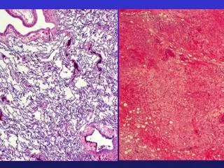
This article discusses the differentiation between pulmonary masses and nodules based on size and etiologic factors. It also provides a classification of etiologic factors in solitary pulmonary masses, including developmental, infectious, neoplastic, inhalational, traumatic, and immunologic causes.

E N D
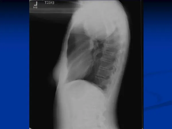
Evaluation of Solitary Lung Mass Vishal Sagar, M.D. Chicago Medical School Chicago, Illinois
Pulmonary Mass vs Pulmonary Nodule • Most authorities consider a size of 3-4 cms as the cut off limit for differentiation between pulmonary mass and pulmonary nodule • Reason for differentiation- different etiologic factors need to be considered if the size is greater than 3-4 cms
Classification of Etiologic factors in a Solitary Pulmonary Mass • Developmental • Infectious • Neoplastic • Inhalational • Traumatic • Immunologic
Developmental • Intralobar sequestration • Almost invariably contiguous to the diaphragm in the posterior bronchopulmonary segment • Typically well defined margin • Cyst formation relatively common • Although cystic in nature, mass remains homogeneous until communication is established with contiguous lung as a result of infection
Infectious Granulomas • Histoplasmosis • Coccidiodomycosis • Tuberculosis • Atypical Mycobacteria • Cryptococcosis • Blastomycosis
Lung Abscess • Predilection for posterior portions of upper or lower lobes • Tends to be round • Ill defined margins when acute but well defined when chronic • No calcification • Cavitation is common • Etiology- usually Staph or anaeobes • Mass may remain unchanged for many weeks
Other Infections • Ascariasis • Pneumocystis Carinii • Aspergilloma • Paragonimus Westermani • Hydatid Cyst
Hydatid Cyst • Causative organism- Echinococcus Granulosus • Predilection for lower lobes • Tends to have bizarre, irregular shape • Calcification may be seen - though extremely rare
Neoplastic • Benign • Malignant • Primary Lung Ca • Metastatic
Benign Tumors • Hamartoma • Lipoma • Fibroma
Characteristics that help determine benign nature of the pulmonary mass • Age less than 35 • Absence of risk factors like smoking or exposure to occupational carcinogens • Small size of the mass ( Size of > 3 cms is associated with an 80% chance of malignancy) • Doubling time < 20 Days or > 400 Days • Certain patterns of calcification • Diffuse • Central • Laminated • Pop corn
Primary Pulmonary Carcinoma • Even though all cell types of lung cancer can present as a solitary peripheral lung mass- it is most commonly seen in adenocarcinoma • Margins tend to be ill defined • Foci of calcification seen on CT in about 5% to 10% of large tumors • Cavitation relatively common
Metastasis • Uncommon for pulmonary metastasis to present as a solitary mass • Tends to be sharply defined and lobulated • Calcification is rare- almost exclusively restricted to metastatic osteogenic sarcoma or choondrosarcoma
Inhalational • Foreign Body Inhalation • Lipid Pneumonia • Silicosis • Coal Workers Pneumoconiosis • Round Atelectasis
Foreign Body Inhalation • Broken fragments of teeth • Food particles • Flowering heads of various grasses • Oral medications • Patients might give a history of recent dental work, general anesthesia or there might be a history of altered consciousness predisposing them to foreign body inhalation/aspiration
Lipid Pneumonia • Inflammatory reaction associated with oil or fat in the alveoli • Usually aspiration of mineral oil used as laxative • Dependent portions of upper and lower lobes • Well defined shape but often has a shaggy outer margin • No calcification seen • CT scan often allows specific diagnosis by demonstrating foci of fat attenuation
Silicosis • Initially involves the periphery of the mid and upper lung zones • Margins may be irregular and somewhat ill defined, simulating pulmonary carcinoma • A background pattern of diffuse silicosis may be apparent • Hilar lymph node enlargement is common and may be associated with “eggshell calcification”
Coal Workers Pneumoconiosis • Marked predilection for upper lobes • Shape- similar to large opacities of silicosis • Calcification generally not seen • May Cavitate • A background of diffuse nodular or reticulonodular shadows is usually evident
Round Atelectasis • Most commonly associated with asbestos exposure • Lower zonal predominance, abuts localized area of pleural thickening • Shape is generally round or oval • No calcification • CT shows the mass abutting a thickened pleura; vessels and bronchi curve toward the periphery of the mass
Traumatic • Pulmonary Hematoma • Usually deep to the point of maximal trauma • It is generally sharply defined, round or oval- • No calcification • No cavitation • Resolution may take several months
Immunologic • Wegners Granulomatosis • Sarcoidosis • Extremely Rare for these to present as solitary lung mass
References • Focal and Multifocal Lung Disease, ScientificAmerican Medicine, IV, 1-19 • Textbook of Respiratory Medicine, 3rd Edition,Murray, Nadel, Mason, Boushey. • Fraser and Pare's Diagnosis of Diseases of the Chest, 4th edition- Fraser, Muller, Colman, Pare. • Textbook of Pulmonary Diseases, Fifth edition- G.L.Baum, Emanuel Wolinsky.