Download
1 / 24
240 likes | 858 Vues
Pancreatitis Definition and Etiology. An acute inflammation process of the pancreas with associated escape of the pancreatic enzyme into surrounding tissue. The primary etiologic factors are alcoholism & biliary tract disease.

E N D
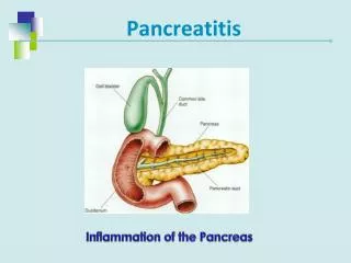
PancreatitisDefinition and Etiology • An acute inflammation process of the pancreas with associated escape of the pancreatic enzyme into surrounding tissue. • The primary etiologic factors are alcoholism & biliary tract disease. • May be a complication of viral or bacterial disease, peptic ulcer, trauma.
PancreatitisIncidence & Risk Factors • Major- Biliary stones, Alcohol use/abuse • Minor- Age: 55 to 65 yrs. for biliary pancreatitis 45- 55 yrs. For alcohol-related • Female for biliary tract pancreatitis; Male-for alcohol-related pancreatitis. • Trauma, Infectious disease, drug toxicities, chronic diseases( inflammatory diseases).
PancreatitisAssessment • Pain: Steady & severe in nature; located in the epigastric or umbilical region; may radiate to the back. Worsened by lying supine; may be lessened by flexed knee, curved-back position. • VomitingVaries in severity, but is usually protracted, worsened by ingestion of food or fluid. Does not relieve the pain. Usually accompanied by nausea.
PancreatitisAssessment con’t…… • Fever: Rarely exceeds 39 C. • Abdominal Finding: Rigidity, tenderness, guarding, distended, decreased or absent peristalsis and paralytic ileus.Fatty stools-(steatorrhea) • Laboratory Finding: Elevation of white count- 20,000-50,000. Elevated serum lipase and amylase(5 to 40 times); glucose, bilirubin, alkaline phosphatase. Urine amylase elevated.Abnormal low serum CA, Na & Mg.-due to dehydration. Binding of Ca in areas of fat necrosis.
PancreatitisNursing Interventions • Alleviate pain & anxiety. Anxiety increases pancreatic secretions. Demerol-then morphine. • Reduce pancreatic stimulus- NPO, NGT to remove gastric secretions. Drugs to reduce pancreatic secretions-anticholinergics-suppress vagal stimulation, NaHco-reverse metabolic acidosis.Regular insulin to treat hyperglycemia. • Prevent or treat infection-with abx. • Aggressive respiratory care- monitor ABG. • Reduce body metabolism- bedrest, cool quiet environment. • Provide client and family instruction-avoid alcohol, coffee,heavy meals and spicy food.
PancreatitisMajor complications • Cardiovascular- hypotension/shock from hypovolemia. • Hematologic-Anemia from blood loss, DIC, leukocytosis from gen.inflammation or secondary infections. • Respiratory-atelectasis, pneumonia, pleural effusion, ARDS • GI- bleeding • Renal- oliguria, acute tubular necrosis • Metabolic-hyperglycemia, hypocalcemia.
CA of the PancreasEtiology • Etiology-unknown. Malignant disease of the exocrine pancreas & more than 85% of the cases are ductal adenocarcinomas. 2/3 develop in the head; remainder occur in the body or tail of the gland. It occurs more commonly in male. • The tumor is usually deeply encased in normal tissue & poorly demarcated. The common duct is often obstructed and distended by the presence of the tumor. Metastasis has almost always occurred before the tumor produces the first symptoms.
CA of the PancreasSigns and Symptoms • Jaundice (lesions of pancreatic head only) • Clay-colored stool • Dark urine • Abdominal pain: usually vague, dull, non-specific • Weight loss • Anorexia • Nausea and vomiting • Glucose intolerance • GI bleeding • Spleenomegaly • ascites
CA of the PancreasInterventions • Non-surgical- High doses of opioid analgesics. Chemotherapy, radiation therapy-intensive external beam radiation therapy by shrinking the tumor cells. • Surgical management: Whipple procedures: the procedure entails the removal of the head of the pancreas, duodenum, a portion of the jejunum, the stomach and the gallbladder, with the anastomosis of the pancreatic duct, the common bile duct, and the stomach to the jejunum.
CA of the PancreasPostoperative Care • Monitor vital parameters. Check vital signs, ABG, intake and output. Be alert to signs of bleeding or shock. Maintain urine output at 30 to 50 ml/hr. • Initiate pulmonary hygiene. • Establish effective pain management. • Monitor dressing and drainage tubes. • Maintain nutritional support with enteral and parenteral support. Monitor BS and insulin. Administer pancreatic enzyme replacement. Assess for signs of dumping syndrome ( rapid shift of fluid from vascular into the intestinal lumen with a resultant decrease in blood volume).
CholelithiasisDefinition, Incidence, Predisposing Factors • Also known as stones in the gallbladder • It is the most common disorder of the biliary system and it has been estimated that 8-10% of all adults in the U.S. have this condition. • Predisposing factors includes: gender, age, estrogen RX or BCP’s, sedentary lifestyle, family history and obesity. • Cholecystitis- inflammation of the gallbladder.
CholelithiasisClinical Manifestations • Sudden-onset pain in the right upper quadrant (RUQ) of the abdomen. Severe and steady in quality. Frequently radiates to the right scapula or shoulder. Persists for abt. 1 to 3 hours. May awaken the patient at night. May be associated with consumption of a large fatty meals. • Anorexia, nausea and vomiting. • Mild to moderate fever • Decreased or absent bowel sounds • Acute abdominal tenderness • Elevated WBC, slightly elevated bilirubin, and alkaline phosphatase.
CholelithiasisDiagnostic Test • Ultrasound-best way to dx; 90-95% effective. • Serum studies- liver function test and serum amylase • Cholangiogram • Gallbladder x-ray test.
CholelithiasisInterventions • Provide relief from vomiting. NGT-reduces distention & eliminates gastric juices that stimulate cholecystokinin. • Maintain fluid and electrolyte balance. • Monitor drug therapy. Administer broad spectrum Abx. Chenodeoxycholic acid- bile acid dissolves cholesterol calculi (60% of the stone). • NTG & papaverine to reduce spasms of duct. • Synthetic narcotics (Demerol, methadone) MSO4 may cause spasms of Oddi and increase spasms.
CholelithiasisInterventions con’t….. • Provide low-fat diet to decrease gallbladder stimulation; avoid alcohol and gas forming foods. • Maintain bedrest • Extracorporeal shock wave lithotripsy- shock wave that disintegrates stones in the biliary system. Ultrasound is used for stone localization before the lithotriptor send waves through a water bag upon which the patient is lying. Analgesics and sedatives to reduce pain during procedures.
CholecystitisAssessment • Epigastric pain- after eating • Pain- localized in RUQ because of somatic sensory nerves. Murphy’s sign- can’t take a deep inspiration when assessor’s fingers are pressed below hepatic margin. Pain begins 2 to 4 hours after eating fried or fatty foods and persist more than 4 to 6 hours. • Nausea, vomiting, anorexia • Low-grade fever • Jaundice • Weight loss
CholecystitisSurgical Management • Cholecystectomy- removal of gallbladder after ligation of the cystic duct and vessels. • Choledochostomy-opening into the common bile duct for removal of stones. T-tube inserted into duct and connected to drainage bottle. Purpose- to decompress biliary tree and allow for postoperative cholangiogram. • Endoscopic cholecystectomy-removal of gallbladder through small puncture hole in the abdomen. Laser dissects gallbladder.
CholecystitisImplementation • Position in low-to semi fowler’s position to facilitate bile drainage. • Maintain skin integrity. • Prevent respiratory complications: TCDB, use of IS. • IF NGT is inserted-to relieve distention and increase peristalsis. • If t-tube inserted-measure amt. & color. Clamp tube before eating. As t-tube clamp-observe for abdominal discomfort and distention. Unclamp if any N/V. • Provide low-fat high carb. and high protein. Maintain for at least 2 to 3 months postoperatively.
Diabetes MellitusDefinition and Classification • Is a chronic disorder of altered CHO, fat & Protein metabolism caused either by: A relative lack of insulin (type 1). Or the inability to respond to insulin (type 11). • Is characterized by persistent hyperglycemia, impaired leukocyte activity & long-term vascular & neurological degeneration
Diabetes MellitusRisk Factors • Familial hx of DM • African-American, Hispanic, or Native American descent. • Obesity • Morbid OB hx. Or hx of delivering infants weighing > 9 lbs.
Diabetes MellitusSignificance • Roughly 7 million people have been dx with DM. • 7th leading underlying cause of death in the U.S. • Leading cause of blindness in adults 20-70 yo. • It accounts for: 30% of new cases of ESRD 50-60% of adult deaths from CAD 40-50% of non traumatic amputations for foot/ankle ulcers.