Download
1 / 1
10 likes | 136 Vues
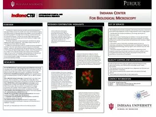
A. B. PT. PT. S1. CL. Bow Sp. PT. PT. D. C. Indiana Center For Biological Microscopy. RESEARCH CONTRIBUTION HIGHLIGHTS. LIST OF SERVICES. OVERVIEW.

E N D
A B PT PT S1 CL Bow Sp PT PT D C Indiana Center For Biological Microscopy RESEARCH CONTRIBUTION HIGHLIGHTS LIST OF SERVICES OVERVIEW • The Imaging Facility provides researchers with training and access to advanced optical microscopy equipment, both for image collection and for image analysis. • Facility personnel provide consultation on the design and analysis of microscopy studies. • The Facility is active in developing new methods of microscopy and digital image analysis, including developing and optimizing multiphoton microscopy, particularly for studies of living animals and developing novel methods of digital image analysis. • The Facility is active in education, providing frequent seminars on microscopy, participating in the graduate program on Biophysical Imaging, the undergraduate Biotechnology training program and a national course for renal researchers. • The Facility maintains a website that in addition to supporting on-line equipment scheduling, also provides numerous tutorials on subjects such as Fluorescence Resonance Energy Transfer, microscopy of living cells and quantification fluorescence co-localization. Funding from Indiana University, the NIH and the Lilly Endowment have given Indiana University a world-class center for biomedical imaging. The Indiana Center for Biological Microscopy (ICBM) is one of a handful in the world equipped for low-light level microscopy, confocal microscopy, UV confocal microscopy, 2-photon microscopy, digital deconvolution microscopy, live cell microscopy and the latest systems for digital image analysis and visualization. The facility represents a strong institutional commitment to optical imaging technology development. Since 1998, Indiana University has committed more than $5M to this facility. In addition to providing state-of-the-art support for School of Medicine members, the core is also actively involved in research into biological imaging, resulting in the development and dissemination of new methods of microscopy and digital image analysis software. One of the products of this research is the ongoing funding of the Indiana University O’Brien Center, whose mission is to develop advanced methods of intravital and 3-dimensional microscopy. The products of these activities are disseminated through a program of education, including seminars, courses and individual training. Volume rendering of a fluorescence microscopy volume of the middle of gastrulating mouse embryo, 7 days into development. The earliest hematopoietic cells (blue) are just emerging from the primitive streak. Michael Ferkowicz and Merv Yoder are looking at these progenitor cells to better understand the proliferation and differentiation of hematopoietic cells during development. 3-dimensional fluorescence image of a colony of endothelial progenitor cells. Nuclei of all cells are labeled with blue Hoechst nuclear stain, and uptake of red acetyl-LDL is used to distinguish early progenitors (center cluster of cells lacking LDL uptake) from spindle cells radiating out from the center and endothelial cells, both of which take up the red fluorescent acetyl-LDL. Daniel Prater and David Ingram are using this cell system to better understand the development of endothelial cells. QUALITY CONTROL AND ASSURANCES RESOURCES • Facility personnel perform rigorous regularly scheduled testing to ensure optimal performance, and modify systems according to users’ needs. • This includes: • Point Spread functions for testing light source alignments and chromatic and spatial aberration of objectives. • Laser power is monitored with a power meter at the plane of the back aperture. • Daily Images with standard slides to test image quality. • Bio-Rad MRC1024 laser scanning confoca and multiphoton microscope is equipped with a Krypton-Argon (488, 568, 647 nm excitations) laser for confocal microscopy and a tunable Titanium-Sapphire laser (using a 5W Millennia diode solid state pump laser) for multiphoton microscopy. • Perkin-Elmer UltraVIEW spinning disk confocal system equipped with 3 lasers providing excitations at 442 nm, 488nm, 514 nm, 568 nm and 647 nm. • Zeiss LSM-510 confocal microscope is equipped with a UV Argon Laser (351 nm, 364 nm excitation), a visible Argon laser (458, 488, 514nm excitation) and two Helium-Neon Lasers (543 nm and 633 nm excitation). • Zeiss LSM510-Meta confocal and multiphoton microscope system equipped with an Argon laser (458, 488, 514nm excitation) and two Helium-Neon Lasers (543 nm and 633 nm excitation) for confocal microscopy, and a tunable Titanium-Sapphire laser (using a 10W Millennia diode solid state pump laser) for multiphoton microscopy. • Inverted Nikon wide-field epifluorescence digital microscope equipped with a Hamamatsu Orca cooled CCD detector. • Upright and inverted Nikon wide-field epifluorescence microscopes equipped with SPOT cooled color CCD detectors. • An Eppendorf micromanipulator and microinjector is available for microinjection studies of living cells • Two Hewlett-Packard workstations devoted to image analysis. • All Microscope Systems and Analysis computers are connected over a 1000Base-T network. • Our staff is an invaluable part of the services that we provide. Intravital multiphoton fluorescence imaging of the bone marrow of the calvarium of a transgenic mouse that expresses GFP in bone marrow hematopoietic progenitors. The vasculature was visualized using a Texas-Red dextran, injected IV 5 minutes prior to imaging. This study is part of a 13 investigator pilot project, led by Nadia Carlesso and Ken Dunn, aimed at developing intravital microscopy as a tool for Cancer Center investigators to study the cancer micro-environment in vivo. This project will culminate in an intravital microscopy service core for the Cancer Center. CONTACT INFORMATION Core Director : Kenneth W. Dunn, Ph.D. Address: Research Institute II, 950 West Walnut St, E233 and E243 Website: http://www.nephrology.iupui.edu/imaging/ Intravital multiphoton fluorescence imaging of filtration in the glomerulus of a living rat. Nucei are visualized with Heochsts (blue). A texas-red dextran was injected after acquisition of frame A. In B the dextran is being filtered from the blood and can be seen within the Bowman’s space of the glomerulus. In C the dextran has been concentrated and appears bright in the distal tubules. In D the dextran has been excreted. This study is part of the O’Brien Center for Advanced Renal Microscopic Analysis, whose primary goal is to develop new optical methodologies for investigators in Nephrologic and Urologic Research.