SGD 1: AMI
SGD 1: AMI. CL, 55y/o M, CEO, severe substernal chest heaviness. SALIENT FEATURES. 55 y/o M, CEO severe substernal chest heaviness accompanied by nausea and vomiting History of having episodes of mild substernal chest tightness for the last 5 yrs

SGD 1: AMI
E N D
Presentation Transcript
SGD 1: AMI CL, 55y/o M, CEO, severe substernal chest heaviness
SALIENT FEATURES • 55 y/o M, CEO • severe substernal chest heaviness • accompanied by nausea and vomiting • History of having episodes of mild substernal chest tightness for the last 5 yrs • a known hypertensive for 6 yrs (Usual BP: 130/80, Highest BP: 170/100) • Irregular intake of Nifedipine 30mg
SALIENT FEATURES • 40 pack years of cigarette smoking • heavy alcohol beverage drinker • Family History • (+ ) premature CAD sister, 38 and brother, 45 • (+ ) HTN father • (+ ) DM mother
SALIENT FEATURES • Soft S1 murmur on PE • Laboratory tests • 12 lead ECG: NSR, ST segment elevation I, aVL, V1-V6; ST segment depression II, III, aVF • Troponin I – 1.5ng/mL • CPK-MB – 256u/L • CXR: cardiomegaly
SALIENT FEATURES • Cont’d • Echo –Doppler: Eccentric LVH w/ segmental wall motion abnormality Ejection fraction at 48% Doppler evidence off impaired LV relaxation Dilated LA Dilated Aortic Root Aortic Sclerosis
Diagnosis Ischemic Cardiac Disease with an Eccentric Left Ventricular Hypertrophy with segmental wall motion abnormality, a Dilated Left Atrium and Aortic Root, and Aortic Sclerosis with evident ST Elevation Myocardial Infarction (STEMI) and Normal Sinus Rhythm, of Class IV-D Functional Disability
Atherogenesis • The process of forming atheromas, plaques in the inner lining (the intima) of arteries. It involves elements of inflammation , a process that provides a unifying theme in the pathogenesis of atherosclerosis
Atherothrombosis • Characterized by an unpredictable, sudden disruption (rupture or erosion/fissure) of an atherosclerotic plaque, which leads to platelet activation and thrombus formation. It is the underlying condition that results in events leading to myocardial infarction, ischemic stroke, and vascular death.
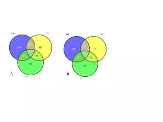
RISK FACTORS OF CAD • Family history of premature CAD • Sex (Male) • Increasing age (>35 years old) • Hyperlipidemia • Hypertension • Stress • Diabetes mellitus • Obesity • Lack of regular exercise • High - fat diet • Cigarette smoking • Too much alcohol
RISK FACTORS PRESENT IN THE PATIENT • (+) Family history of premature CAD (sister at age 38 and brother at age 45) • Sex (Male) • Age (55 years old) • Known hypertensive for 6 years • Highest BP = 170/100 mmHg • Irregular intake of Nifedipine 30 mg • Forty (40) pack years of cigarette smoking • Stress • Occupation: CEO • Had an argument during regular board meeting • Heavy alcoholic beverage drinker
4. What are the WHO criteria for acute myocardial infarction? Are they present in this patient?
WHO CRITERIA FOR AMI (1979): A patient is diagnosed with myocardial infarction if two (probable) or three (definite) of the following criteria are satisfied: 1. Clinical history of ischemic type of chest pain lasting for more than 20 minutes.
WHO CRITERIA FOR AMI (1979): A patient is diagnosed with myocardial infarction if two (probable) or three (definite) of the following criteria are satisfied: 2. Changes in serial ECG tracings.
WHO CRITERIA FOR AMI (1979): A patient is diagnosed with myocardial infarction if two (probable) or three (definite) of the following criteria are satisfied: 3. Rise and fall of serum cardiac biomarkers such as creatininekinase-MB fraction and troponin.
REDEFINED WHO CRITERIA FOR AMI (2000): The WHO criteria were refined in 2000 to give more prominence to cardiac biomarkers. Cardiac troponin rise accompanied by either: Typical symptoms Pathological Q waves ST elevation or depression Coronary intervention
IN THE PATIENT: CRITERIA: • Severe substernal heaviness (experienced about 1 hour) • ST segment elevation I, aVL, V1-V6 • Troponin I: 1.5ng/mL • (NV: 0 – 0.4 ng/mL) • CPK-MB: 256 u/L • (NV: <1 u/L) • ST segment depression II, III and aVF • Clinical history of ischemic type chest pain lasting > 20 mins • Changes in serial ECG tracings • Rise and fall of cardiac biomarkers • Cardiac troponin rise accompanied by ST depression
5.What are the clinical features(Sx and PE) of AMI? Are they present in this patient?
Symptoms and PE of AMI • Chest pain • described as a pressure sensation, fullness, or squeezing in the midportion of the thorax • may radiate to the arms, jaw or teeth, shoulder, and/or back • Associated diaphoresis or sweating palpitations, weakness Substernal chest pain for >30 mins and diaphoresis strongly suggests STEMI • Presence of a precipitating factor: • Vigorous physical activity • Emotional stress • Illness: medical or surgical
Symptoms and PE of AMI • Associated with dyspnea or shortness of breath • Associated epigastric discomfort with or without nausea and vomiting • Syncope or near-syncope without other cause • Impairment of cognitive function without other cause lightheadedness or loss of consciousness a look of anxiety, or a sense of impending doom or death • It may occur at any time of the day, but most appear to be clustered around the early hours of the morning, are associated with demanding physical activity, or both
Symptoms and PE of AMI PE • Signs of ventricular dysfunction: • (+) S3 and S4 • Decreased intensity of S1 • Paradoxical splitting of S2 • Transient midsystolic or late systolic apical systolic murmur (mitral valve apparatus dysfunction) • Anterior infraction (1/4 of pxs): sympathetic hyperactivity • Tachycardia, hypertension • Posterior infarction (1/2 of pxs): parasympathetic hyperactivity • Bradycardia, hypotension • Carotid pulse decreased in volume (reduced stroked volume) • 1st week after STEMI: temp. up to 38 ⁰C • in transmural STEMI: (+) pericardial friction rub
Symptoms and PE seen in AMI which is present in our patient • (+) precipitating factor • Emotional stress: started after argument during board meeting • (+) Chest pain (severe substernal chest heaviness) not relieved by rest • (+) weakness, sweating ( due to sympathetic activity), nausea, vomiting anxiety • Soft S1
STEMI ECG FINDINGS ST-segment elevation of at least 1 mm in two or more limb leads
STEMI ECG FINDINGS At least 2 mm ST segment elevation in two or more precordial leads
STEMI • ST segment elevation • Q – wave inversion
ECG NSTEMI • In UA, • ST-segment depression • transient ST-segment elevation • T-wave inversion
Ischemia Infarction Fibrosis Non-ST Elevation Infarction ST depression & T-wave inversion The ECG changes seen with a non-ST elevation infarction are: Before injury Normal ECG ST depression & T-wave inversion ST returns to baseline, but T-wave inversion persists
What is the location of the AMI in this patient based on his ECG?
ECG of the Patient • ST segment depression on II, III and aVF • Marked ST elevation on I, aVL, V1-V6
Early Management of AMI Key points • Recognition of symptoms by the patient and prompt seeking of medical attention • Rapid deployment of emergency medical team capable of performing resuscitative measures • Expeditious transport of patient to hospital • Early treatment without more delay
Early Management of AMI Goals of Management • Control of cardiac discomfort • Rapid identification of patients in need of urgent reperfusion therapy
Early Management of AMI Treatment: 1. Aspirin- rapidly inhibits COX-1 in platelets and reduces thromboxane A2. - chewed 160- 325 mg tablet 2. Presence of hypoxemia- Oxygen supplementation (2-4 L/ min) 3. Nitroglycerin(sublingual) – 3 doses of 0.4 mg at 5 min intervals to relieve chest pain and decrease preload and dilate infarcted coronary or collateral vessels.
Early Management of AMI 4. Nitroglycerin (intravenous)- given when there is return of chest pain accompanied with ST-segment of T-wave shifts. 5. Beta-blockers- diminish myocardial O2 demand and ischemia, reduces the risk of ventricular fibrillation. 6. Reperfusion therapy- by fibrinolysis or primary Percutaneous Coronary Intervention. - done if ST- segment elevation is at least 2mm in 2 contiguous precordial leads and 1 mm in 2 adjacent limb leads present
Complications • Ventricular remodeling • left ventricle undergoes a series of changes in shape, size, and thickness in both the infarcted and noninfarcted segments • Precedes clinically evident CHF in months to years after infarction • Progressive dilation and its clinical consequences may be ameliorated by therapy with ACE inhibitors and other vasodilators (e.g., nitrates). • Ventricular premature beats • Infrequent, sporadic ventricular premature depolarizations occur in almost all patients with STEMI and do not require therapy. • hypokalemia and hypomagnesemia are risk factors for ventricular fibrillation in patients with STEMI; the serum potassium concentration should be adjusted to approximately 4.5 mmol/L and magnesium to about 2.0 mmol/L.
Complications • Ventricular tachycardia and fibrillations • Within the first 24 h of STEMI, ventricular tachycardia and fibrillation can occur without prior warning arrhythmias. • The occurrence of ventricular fibrillation can be reduced by prophylactic administration of intravenous lidocaine. • Ventricular arrhythmias, including the unusual form of ventricular tachycardia known as torsades des pointes, may occur in patients with STEMI as a consequence of other concurrent problems (such as hypoxia, hypokalemia, or other electrolyte disturbances) or of the toxic effects of an agent being administered to the patient (such as digoxin or quinidine)
Complications • Accelerated idioventricular rhythm (AIVR, "slow ventricular tachycardia") • Ventricular rhythm with a rate of 60–100 bpm, occurs in 25% of patients with STEMI. • Often occurs transiently during fibrinolytic therapy at the time of reperfusion. • Benign and does not presage the development of classic ventricular tachycardia. • Most episodes do not require treatment if the patient is monitored carefully, as degeneration into a more serious arrhythmia is rare. • Sinus bradycardia • Treatment of sinus bradycardia is indicated if hemodynamic compromise results from the slow heart rate. Atropine is the most useful drug for increasing heart rate and should be given intravenously in doses of 0.5 mg initially.
Complications • Recurrent chest discomfort • Recurrent angina develops in ~25% of patients hospitalized for STEMI. • Higher in patients who undergo successful fibrinolysis. • Since recurrent or persistent ischemia often heralds extension of the original infarct or reinfarction in a new myocardial zone and is associated with a near tripling of mortality after STEMI, patients with these symptoms should be referred for prompt coronary arteriography and mechanical revascularization. • Repeat administration of a fibrinolytic agent is an alternative to early mechanical revascularization.