Understanding the Muscular System: Types, Functions, and Mechanics
490 likes | 610 Vues
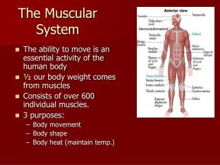
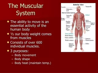
The muscular system encompasses fibrous tissues capable of contraction, essential for movement, posture maintenance, and critical bodily functions such as heartbeat regulation and fluid propulsion. It consists of three types of muscles: skeletal for voluntary movement, smooth for involuntary processes like digestion, and cardiac for heart function. Structure-wise, skeletal muscles feature layers of connective tissue and specialized fibers that react to neural stimulation, leading to muscle contractions. Energy for these contractions is derived from ATP and oxygen, with insights into mechanisms like the sliding filament model and muscle fatigue.

Understanding the Muscular System: Types, Functions, and Mechanics
E N D
Presentation Transcript
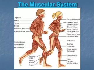
The Muscular System By: Kendra Iverson & Sarah Bender
What does it means? • Muscle- fibrous tissue with the ability to contract, producing movement in or maintaining the position of an animal body • System- organized arrangement; network …. • Set of connected fibrous tissue that work together to produce movement
Functions of Muscles • Provide muscle tone • Propel body fluids and food • Generate the heartbeat • Distribute heat throughout the body
Types of Muscles • Skeletal muscle • Moves majority of the body • Smooth muscle • Helps most with digestive system • Cardiac muscle • Controls the heartbeat
Skeletal Muscle • Location: all skeletal muscles- attach to bones • Function: movement of bones at joints and maintaining posture • Voluntary • Contracts and relaxes quickly • Transverse tubule system is well-developed
Skeletal Muscle Structure • Composed of: • skeletal muscle tissue • nervous tissue • blood • connective tissue
Skeletal Muscle Tissues • Fascia- layers of fibrous connective tissue • covers and separates each muscle surface • Epimysium- lies beneath the fascia • Perimysium- surrounds individual bundles of fibers (fascicles) • Endomysium- connective tissue layer that separates individual muscle fibers
Skeletal Muscle Fibers • Cells that contract in response to stimulation and then relax after the stimulation ends • Thin and elongated cylinders with rounded ends • Can extend full length of a muscle
(Continued) Details • Sarcolemma- muscle fiber membrane ↓ • Sarcoplasm- cytoplasm ↓ • Myofibrils- protein filaments which lie parallel and play great role in contraction • Types: Myosin- thicker, called A bands (dark) Actin- thinner, called I bands (light) ↓ • Sarcomere- space from center of one I band to the next I band
(Continued) Details • Sarcoplasmic Reticulum- network of membranous channels that surround each myofibril and run parellel • Transverse Tubules (T tubules)- network of membranous channels between sarcoplasm that open outside • Both- activate muscle contraction mechanism when the fiber is stimulated
Neuromuscular Junction • Site where the motor neuron and muscle fiber meet • Muscle fiber is specialized to form a motor end plate • End branches, has many mitochondria and contains synaptic vesicles that store chemicals • Neurotransmitter stimulates muscle fibers to release acetylcholine • Travels down a motor neuron axon and stimulates muscle fiber contraction
Motor Unit • Includes one motor neuron and several muscle fibers • The motor neuron transmits an impulse, which allows all the linked muscle fibers to contract at the same time
Skeletal Muscle Contraction • Sarcomeres shorten and the muscle is pulled against its attachments • Sliding Filament Model • Myosin cross-bridge attaches to the binding site on the actin filament and bends, causing a pull on the actin filament • Myosin releases and attaches to the next binding site on the actin, causing a pull • ATP converts to ADP • Repeats this cycle as long as the energy source ATP is available and the muscle fiber is stimulated to contract
Stimulus for Contraction • Stimulated by acetylcholine (neurotransmitter) • Motor neuron releases acetylcholine into the synaptic cleft to initiate contraction • Motor end plate has protein receptors which detect acetylcholine • Muscle impulse travels over sarcolemma surface and into T tubules, reaching the sarcoplasmic reticulum
Stimulus for Contraction • Stored calcium ions in the sarcoplasmic reticulum get released into the sarcoplasm • Troponin and tropomyosin (proteins associated with actin) move to expose the myosin binding sites on the actin filaments due to the concentration of calcium • Muscle shortens because myosin binds and pulls on actin • After received- acetylcholinesterase breaks down acetylcholine, calcium returns to sarcoplasmic reticulum, and myosin and actin links break
Energy Sources for Contraction • ATP molecules provides the energy • Must be regenerated because it has a limited supply • Creatine phosphate • Energy can be transferred to ADP molecules, converting them back ATP molecules as ATP is decomposing
Oxygen Supply & Cellular Respiration • Cellular respiration makes a few ATP molecules in early phases • Muscles require oxygen • Glucose in broken down completely in mitochondria • Blood carries oxygen, bound to the pigment hemoglobin • The pigment myoglobin stores the oxygen in muscle tissue
Oxygen Debt • The amount of oxygen that liver cells need to convert lactic acid into glucose, and the amount muscle cells must have to recreate the original concentration of ATP and creatine phosphate • Develops during strenuous activity • Pyruvic acid builds up during anaerobic respiration and diffuses out of muscle cells into the liver • May take hours to repay
Muscle Fatigue • When a muscle can no longer contract • Usually from a build up of lactic acid in the muscle, which lowers pH and results in muscle fibers to not respond to stimulation • Cramps- a muscle has a sustained involuntary contraction
Heat Production • More than half of energy released in cellular respiration becomes heat • All active cells generate heat, but muscle cells make up a large majority of a bodies total mass • Blood moves heat from muscle to other tissues, maintaining body temperature
Smooth Muscle • Location: walls of hollow internal organs, blood vessels • Function: to move food through the digestive system and constrict blood vessels • Involuntary • Contracts and relaxes slowly • Rhythmic • No transverse tubules
Smooth Muscle Fibers • Major types: • Multiunit: muscle fibers are separate • Usually contract in response to stimulation by certain hormones or motor nerve impulses • Visceral: organized in sheets • Are found in walls of hollow organs • Can stimulate others, displaying rhythmically • Responsible for involuntary wavelike motion called peristalsis
Smooth Muscle Contraction • Reactions of actin and myosin • Triggered by membrane impulses and increased intracellular calcium ions • Uses energy from ATP • Neurotransmitters are acetylcholine and norepinephrine • Affected by hormones • Fibers can change length without changing tension
Cardiac Muscle • Location: wall of the heart ONLY • Function: pumping action of the heart • Involuntary • Network of cells contract as a unit • Rhythmic • Transverse tubule system is well-developed • Intercalated discs separating adjacent cells
Cardiac Muscle Contraction • Intercalated discs communicate a muscle impulse throughout entire heart • Allows the hearts muscle fibers to beat as a single unit • Network responds in an all-or-none manner
Muscular Responses • Threshold Stimulus- the minimal strength required to cause a contraction • Muscle fibers remain unresponsive until reached
Muscular Responses • All-or-None Response- if a muscle fiber contracts at all, it contracts fully • There is no partial contraction • Once threshold stimulus is reached, fiber responds to fullest contraction
Muscular Responses • Recording a Muscle Contraction: • Myogram- the pattern resulting from a movement when connected to a device • Twitch- a single contraction that lasts only a fraction of a second • Latent period- the delay between the time of stimulation and the time the fiber responds
(a) Series of twitches (b) Summation (c) Tetanic Contraction
Muscular Responses • Summation- when the stimulus of the next series arrives before the muscle fiber can completely relax • Tetanic Contraction- when a forceful, sustained contraction lacks any relaxation
Muscular Responses • Recruitment of Motor Units: • Recruitment- the number of motor units activated as a result of a higher intensity of stimulation • The addiction of muscle fibers to take part in a contraction
Muscular Responses • Sustained Contractions: • Produced by summation and recruitment together • Increasing strength • Muscle tone- fibers undergo some sustained contraction even when muscles appear to be relaxed • Keeps posture maintained
Use & Disuse of Skeletal Muscles • Muscular hypertrophy: muscles enlarging in size and strength due to forceful exercise • Muscular atrophy: muscles decreasing in size and strength due to lack of exercise • Size is relative to strength • Capillary networks, actin and myosin filaments, and number of mitochondria
Use & Disuse of Skeletal Muscles • Slow twitch fibers: lower intensity and able to resist fatigue, but may not increase muscle size • Running or swimming • Fast twitch fibers: higher intensity and increase muscle size, but are fatigable • Lifting weights
Skeletal Muscle Actions • Variety of movements • Movement depends on: • The kind of joint • How the muscle attaches
Origin & Insertion • Origin: the immovable end of a muscle • Insertion: the movable end of a muscle • The insertion is pulled towards the origin when contraction occurs • Flexion and extension tell about the angle between bones that meet at a joint
Interaction of Skeletal Muscles • Almost always function in groups • Must will movement to occur and nervous system allows correct muscles to stimulate • Prime Mover/Agonist: muscle responsible for a particular movement • Synergist: muscles that contract and assist the prime mover • Antagonists: muscles that resist the action of the prime mover
Disorders/Diseases • Tendinitis- a tendon (attaches muscle to bone) becomes inflamed and swollen after injury or repeated stress during activity • Muscle strain- connective tissues are overstretched and the degree of damage and seriousness varies
Disorders/Diseases • Botulinum toxin- blocks stimulation of muscle fibers, paralyzing muscles • “Botox” to temporarily smooth wrinkles by not allowing to move facial muscles • Steroids- (+) increase muscular strength and used for medical purposes, (-) cause changes of opposite sex and damage internal organs
Inherited Diseases • Muscular Dystrophies • Missing proteins • Muscles weaken and degenerate • Charcot-Marie-Tooth Disease • A duplicate gene • Weakens hands and feet, and tendon reflexes • Myotonic Dystrophy • An extending gene • Muscle weakness and irregular heartbeats • Hereditary Idiopathic Dilated Cardiomyopathy • A glitch • Rare form of heart failure in actin