Glycogen metabolism
Glycogen metabolism. UNIT II: Intermediary Metabolism. Figure 11.1. Glycogen synthesis and degradation shown as a part of the essential reactions of energy metabolism (see Figure 8.2, p. 90, for a more detailed view of the overall reactions of metabolism). Overview.

Glycogen metabolism
E N D
Presentation Transcript
Glycogen metabolism UNIT II: Intermediary Metabolism
Figure 11.1. Glycogen synthesis and degradation shown as a part of the essential reactions of energy metabolism (see Figure 8.2, p. 90, for a more detailed view of the overall reactions of metabolism)..
Overview • A constant source of blood glucose is an absolute requirement for human life • Glucose is the greatly preferred energy source for the brain, & the required energy source for cells with few or no mitoch., e.g., mature RBCs • Glucose is also essential as an energy source for exercising muscle, where it is the substrate for anaerobic glycolysis. • Blood glucose can be obtained from 3 primary sources: diet, degradation of glycogen, & gluconeogenesis. • Dietary intake of gluc & gluc precursors, e.g., starch, monosacch’s, & disacch’s, is sporadic and, depending on diet, is not always a reliable source of gluc. • In contrast, gluconeogenesis can provide sustained synthesis of gluc, but it is somewhat slow in responding to falling blood gluc level • Therefore, body has developed mechanisms of storing a supply of glucose in a rapidly mobilizable form i.e., glycogen.
In absence of a dietary source of gluc, this cpd is rapidly released from liver & kidney glycogen. Similarly, muscle glycogen is extensively degraded in exercising muscle to provide that tissue with an important energy source • When glycogen stores are depleted, specific tissues synthesize gluc de novo, using aa’s from body’s proteins as primary source of carbons for gluconeogenic pathway.
II. Structure and function of glycogen • The main stores of glycogen in the body are found in skeletal muscle & liver, although most other cells store small amounts of glycogen for their own use • Function of muscle glycogen is to serve as a fuel reserve for synthesis of ATP during muscle contraction • That of liver glycogen is to maintain blood glucose conc., particularly during early stages of fast A. Amounts of liver and muscle glycogen • ~ 400 g of glycogen make up one to two percent of the fresh weight of resting muscle, & ~ 100 g of glycogen make up to 10% of the fresh weight of a well-fed adult liver. What limits production of glycogen at these levels is not clear. • However, in some glycogen storage diseases, the amount of glycogen in liver and/or muscle can be significantly higher
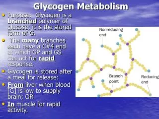
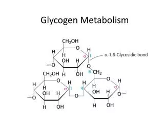
B. Structure of glycogen • Glycogen is a branched-chain homo-polysaccharide made exclusively from α-glucose • The primary glycosidic bond is an α(1→4) linkage. • After an av. of 8-10 glucosyl residues, there is a branch containing an α(1→6) linkage. • A single molecule of glycogen can have a molecular mass of up to 108 daltons. • These molecules exist in discrete cytoplasmic granules that contain most of the enz’s necessary for glycogen synthesis & degradation
Figure 11.3. Branched structure of glycogen, showing a-1,4 and a-1,6 linkages.
C. Fluctuation of glycogen stores • Liver glycogen stores increase during the well-fed state & are depleted during a fast • Muscle glycogen is not affected by short periods of fasting ( a few days) & is only moderately decreased in prolonged fasting (weeks). • Muscle glycogen is synthesized to replenish muscle stores after they have been depleted, e.g., following strenuous exercise. Note: synthesis & degradation of glycogen are processes that go on continuously. Differences b/w rates of these 2 processes determine levels of stored glycogen during specific physiologic states
III. Synthesis of glycogen (glycogenesis) - Glycogen is synthesized from molecules of α-D-glucose. The process occurs in cytosol, and requires energy supplied by ATP (for phosphorylation of gluc) & uridine triphosphate (UTP) A. Synthesis of UDP-glucose • α-D-gluc attached to UDP is the source of all of glucosyl residues that are added to the growing glycogen molecule • UDP-gluc is synthesized from glucose-1-P & UTP by UDP-glucose pyrophosphorylase • The high-energy bond in pyrophosphate (PPi), the 2nd product of the reaction, is hydrolyzed to 2 inorganic phosphates (Pi) by pyrophosphatase, which ensures that synthesis of UDP-gluc proceeds in direction of UDP-gluc production Note: G-6-P is converted to G-1-P by phosphoglucomutase. G-1,6-BP is an obligatory intermediate in this reaction
B. Synthesis of a primer to initiate glycogen synthesis • Glycogen synthase is responsible for making α (1→4) linkages in glycogen. This enz can’t initiate chain synthesis using free gluc as an acceptor of a molecule of gluc from UDP-gluc. Instead, it can only elongate already existing chains of gluc • Therefore, a fragment of glycogen can serve as a primer in cells whose glycogen stores are not totally depleted • In the absence of a glycogen fragment, a protein, called glycogenin, can serve as an acceptor of gluc residues • Side chain hydroxyl group of a specific Tyr serves as the site at which the initial glucosyl unit is attached • Transfer of first few molecules of gluc from UDP-gluc to glycogenin is catalyzed by glycogenin itself, which can then transfer additional glucosyl units to the growing α (1→4)-linked glucosyl chain • This short chain serves as an acceptor of future gluc residues Note: glycogenin stays associated with & is found in center of completed glycogen molecule
C. Elongation of glycogen chain by glycogen synthase - Elongation of glycogen chain involves transfer of gluc from UDP-gluc to the non-reducing end of growing chain, forming a new glycosidic bond b/w the anomeric hydroxyl of C-1 of activated gluc & C-4 of accepting glucosyl residue Note: “non-reducing end” of a CHO chain is one in which anomeric C of terminal sugar is linked by a glycosidic bond to another cpd, making terminal sugar “non-reducing”. - The enz responsible for making α (1→4) linkages in glycogen is glycogen synthase Note: UDP released when the new α (1→4) glycosidic bond is made can be converted back to UTP by nucleoside diphosphate kinase (UDP + ATP ↔ UTP + ADP)
Figure 11.6. Interconversion of glucose 6-phosphate and glucose 1-phosphate by phosphoglucomutase.
D. Formation of branches in glycogen • If no other synthetic enz’s acted on the chain, resulting structure would be a linear molecule of glucosyl residues attached by α (1→4) linkages. • Such a cpd is found in plant tissues, & is called amylose. In contrast, glycogen has branches located, on av., 8 glucosyl residues apart, resulting in a highly branched, tree-like structure that is far more soluble than unbranched amylose • Branching also increases the # of non-reducing ends to which new glucosyl residues can be added (and also, from which these residues can be removed), thereby greatly accelerating the rate at which glycogen synthesis & degradation can occur, & dramatically increasing the size of the molecule
1. Synthesis of branches: • Branches are made by action of “branching enzyme”, amylo-α (1→4) → α (1→6)-transglucosidase. This enz transfers a chain of 5 to 8 glucosyl residues from non-reducing end of glycogen chain [breaking α (1→4) bond] to another residue on the chain and attaches it by an α (1→6) linkage • Resulting new, non-reducing end, as well as the old non-reducing end from which the 5 to 8 residues were removed, can now be elongated by glycogen synthase 2. Synthesis of additional branches: - After elongation of these two ends has been accomplished by glycogen synthase, their terminal 5 to 8 glucosyl residues can be removed & used to make further branches
IV. Degradation of glycogen (glycogenolysis) • The degradative pathway that mobilizes stored glycogen in liver & skeletal muscle is not a reversal of the synthetic reactions. Instead a separate set of cytosolic enz’s is required. • When glycogen is degraded, the primary product is G-1-P, obtained by breaking α (1→4) glycosidic bonds. In addition, free gluc is released from each α (1→6)-linked glucosyl residue
A. Shortening of chains • Glycogen phosphorylase sequentially cleaves the α (1→4) glycosidic bonds b/w the glucosyl residues at the non-reducing ends of glycogen chains by simple phosphorolysis until 4 glucosyl units remain on each chain before a branch point Note: this enz contains a molecule of covalently bound pyridoxal phosphate that is required as a coenzyme - Resulting structure is called a limit dextrin, & phosphorylase can’t degrade it any further
B. Removal of branches • Branches are removed by 2 enzymatic activities. 1stoligo-α(1→4)→α (1→4)-glucan transferase removes the outer 3 of the 4 glucosyl residues attached at a branch. It next transfers them to the non-reducing end of another chain, lengthening it accordingly. Thus, an α(1→4) bond is broken and an α(1→4) bond is made. • Next, the remaining single gluc residue attached in an α(1→6) linkage is removed hydrollytically by amylo- α(1→6)-glucosidase activity, releasing free gluc. Note: both the transferase & glucosidase are domains of a single polyp molecule, the ‘debranching enzyme”. - The glucosyl chain is now available for degradation by glycogen phosphorylase until 4 glucosyl units from next branch are reached
Figure 11.8 Glycogen degradation, showing some of the glycogen storage diseases. (Continued on next page.)
C. Conversion of G-1-P to G-6-P • G-1-P, produced by glycogen phosphorylase, is converted in the cytosol to G-6-P by phosphoglucomutase, a reaction that produces G-1,6-BP as a temporary but essential intermediate • In liver, G-6-P is translocated into ER by glucose 6-phosphate translocase. There it is converted to glucose by glucose 6-phosphatase, the same enz used in last step of gluconeogenesis • Resulting glu is then transported out of ER to cytosol. Hepatocytes release glycogen-derived gluc into blood to help maintain blood gluc levels until gluconogenic pathway is actively producing gluc Note: in muscle, G-6-P can’t be dephosphorylated because of a lack of glucose-6-phosphatase. Instead, it enters glycolysis, providing energy needed for muscle contraction
D. Lysosomal degradation of glycogen • A small amount of glycogen is continuously degraded by lysosomal enz, α(1→4)-glucosidase (acid maltase). Purpose of this pathway is unknown • However, a deficiency of this enz causes accumulation of glycogen in vacuoles in the cytosol, resulting in the serious glycogen storage disease type II (Pompe disease)
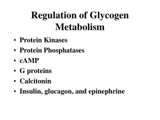
V. Regulation of glycogen synthesis & degradation • Because of importance of maintaining blood gluc levels, synthesis & degradation of its glycogen storage form are tightly regulated • In liver, glycogen synthesis accelerates during periods when the body has been well fed, whereas degradation accelerates during periods of fasting. • In skeletal muscle, glycogen degradation occurs during active exercise, & synthesis begins as soon as the muscle is again at rest • Regulation of glycogen synthesis & degradation is accomplished on two levels
First, glycogen synthase & glycogen phosphorylase are allosterically controlled. • Second, the pathways of glycogen synthesis & degradation are hormonally regulated Note: regulation of glycogen synthesis & degradation is extremely complex, involving many enz’s (e.g., protein kinases & phosphatases), calcium, & enz inhibitors, among others
A. Allosteric regulation of glycogen synthesis & degradation • Glycogen synthase & glycogen phosphorylase respond to levels of metabolites & energy needs of cell. It is logical, therefore, that glycogen synthesis is stimulated when substrate availability & energy levels are high, whereas glycogen degradation is increased when energy levels & available gluc supplies are low 1. Regulation of glycogen synthesis & degradation in the well fed state: • In the well fed state, glycogen synthase is allosterically activated by G-6-P when it is present in elevated conc’s. in contrast, glycogen phosphorylase is allosterically inhibited by G-6-P, as well as ATP, a high-energy signal in cell. Note: in liver, gluc also serves as an allosteric inhibitor of glycogen phosphorylase
Figure 11.9. Allosteric regulation of glycogen synthesis and degradation in A. Liver, and B. Muscle.
2. Activation of glycogen degradation in muscle by calcium • During muscle contraction, there is a rapid & urgent need for ATP, the energy for which is supplied by muscle’s stores of glycogen. • Nerve impulses cause memb depolarization, which in turn promotes Ca2+ release from sarcoplasmic reticulum into sarcoplasm of muscle cells • Ca2+ binds to calmodulin, one of a family of small, calcium-binding proteins Note: calmodulin is the most widely distributed of these proteins, & is present in virtually all cells - Binding of 4 molecules of Ca2+ to calmodulin triggers conformational change such that activated Ca2+-calmodulin complex binds to & activates protein molecules, often enz’s, that are inactive in absence of this complex • Thus, calmodulin functions as an essential subunit of many complex proteins. One such protein is phosphorylase kinase, which is activated by Ca2+-calmodulin complex without need for the kinase to be phosphorylated by cAMP-dependent protein kinase • When muscle relaxes, Ca2+ returns to sarcoplasmic reticulum & phosphorylase kinase becomes inactive Note: phosphorylase kinase is maximally active in exercising muscle when it is both phosphorylated & bound to Ca2+
Figure 11.10. Calmodulin mediates many effects of intracellular calcium.
3. Activation of glycogen degradation in muscle by AMP • Muscle glycogen phosphorylase is active in presence of high AMP conc’s that occur in muscle under extreme conditions of anoxia & ATP depletion • AMP binds to the inactive form of glycogen phosphorylase, causing its activation without phosphorylation
B. Activation of glycogen degradation by cAMP-directed pathway • Binding of hormones, e.g., glucagon & epinephrine, to memb receptors signals the need for glycogen to be degraded, either to elevate blood gluc levels or to provide energy for exercising muscle 1. Activation of protein kinase: • Binding of glucagon or epinephrine to their specific CM receptors cAMP-mediated activation of cAMP-dependent protein kinase. • This enz is a tetramer, having 2 regulatory (R) & 2 catalytic (C) subunits. • cAMP binds R subunit dimer, releasing individual C subunits that are active Note: when cAMP removed, inactive tetramer R2C2, is again formed
2. Activation of phosphorylase kinase: • Phosphorylase kinase exists in 2 forms: an inactive “b” form & an active “a” form • Active cAMP-dependent protein kinase phosphorylates inactive form of phosphorylase kinase activation Note: phosphorylated enz can be inactivated by hydrolytic removal of its P by protein phosphatase 1. This enz is activated by a kinase-mediated signal cascade initiated by insulin 3. Activation of glycogen phosphorylase: • Glycogen phosphorylase also exists in 2 forms: the dephosphorylated, inactive “b” form & phosphorylated, active “a” form. • Active phosphorylase kinase phosphorylates glycogen phosphorylase a, which then begins glycogen breakdown
- Phosphorylase a is converted to phosphorylase b by hydrolysis of its P by protein phosphatase 1. Note: - when gluc is bound to glycogen phosphorylase a, thus signaling that glycogen degradation is no longer required, the complex becomes a better substrate for protein phosphatase 1. • In addition, when muscle glycogen phosphorylase b is bound to glucose, it can’t be allosterically activated by AMP • In the muscle, insulin indirectly inhibits the enz by increasing uptake of gluc, leading to an increased level of G-6-P, a potent allosteric inhibitor of glycogen phosphorylase
Figure 11.11. Stimulation and inhibition of glycogen degradation.
4. Summary of regulation of glycogen degradation: • Cascade of reactions listed above result in glycogen degradation • The large # of sequential steps serves to amplify the effect of the hormonal signal, i.e., a few hormone molecules binding to their receptors result in a # of protein kinase molecules being activated that can each activate many phosphorylase kinase molecules • This causes production of many active glycogen phosphorylase a molecules that can degrade glycogen
C. Inhibition of glycogen synthesis by a cAMP-directed pathway • The regulated enz in glycogen synthesis is glycogen synthase. • It also exists in 2 forms, the “a” form which is not phosphorylated & is the most active form, & “b” form, which is phosphorylated & inactive. • Glycogen synthase a is converted to “b” form by phosphorylation at several sites on the enz, with the level of inactivation is proportional to its degree of phosphorylation • This conversion process is catalyzed by several different protein kinases that are regulated by cAMP or other signaling mechanisms Note: protein kinase C, a Ca2+ & phospholipid-dependent protein kinase, also phosphorylates glycogen synthase. Neither protein kinase A nor C directly phosphorylates glycogen phosphorylase
- Binding of glucagon or epinephrine to hepatocyte receptors, or of epinephrine to muscle cell receptors, results in the activation of adenylyl cyclase, mediated by G-protein • This enz catalyzes synthesis of cAMP, which activates cAMP-dependent protein kinase A • Protein kinase A then phosphorylates & thereby inactivates glycogen synthase • Glycogen synthase “b” can be transformed back to synthase “a” by protein phosphatase 1, which removes P groups hydrolytically
Figure 11.12. Hormonal regulation of glycogen synthesis. [Note: In contrast to glycogen phosphorylase, glycogen synthase is inactive if phosphorylated.]
VI. Glycogen storage diseases • These are a group of genetic diseases that result from a defect in an enz required for glycogen synthesis or degradation • They result either in formation of glycogen that has an abnormal structure, or in the accumulation of excessive amounts of normal glycogen in specific tissues as a result of impaired degradation • A particular enz may be defective in a single tissue, such as liver, or the defect may be more generalized, affecting liver, muscle, kidney, intestine, & myocardium • Severity of glycogen storage diseases (GSDs) ranges from fatal in infancy to mild disorders that are not life-threatening
Summary • Main stores of glycogen in body are found in skeletal muscle, where they serve as a fuel reserve for synthesis of ATP during muscle contraction, & in liver, where glycogen is used to maintain blood glucose conc, particularly during early stages of a fast • Glycogen is a highly branched polymer of α-D-glucose. The primary glycosidic bond is an α (1→4) linkage. After ~ 8-10 gluc residues, there is a branch containing an α (1→6) linkage. • UDP-gluc, building block of glycogen, is synthesized from G-1-P & UTP by UDP-glucose pyrophosphorylase • Gluc from UDP-glucose is transferred to the non-reducing ends of glycogen chains by glycogen synthase, which makes α (1→4) linkages
Branches are formed by amylo-α(1→4) → α(1→6)-transglucosidase, which transfers a chain of 5-8 glucosyl residues from the non-reducing end of glycogen chain (breaking an α(1→4) linkage), and attaches it with an α(1→6) linkage to another residue in the chain • Glycogen phosphorylase cleaves the α(1→4) bonds b/w glucosyl residues at the non-reducing ends of glycogen chains, producing G-1-P. it requires pyridoxyl phosphate as a coenz. • This sequential degradation continues until 4 glucosyl units remain on each chain before a branch point. The resulting structure is called a limit dextrin. • Oligo-α(1→4)→α(1→4)-glucan transferase [common name, glucosyl (4:4) transferase] removes the outer 3 of the 4 glucosyl residues attached at a branch, & transfers them to the non-reducing end of another chain where they can be converted to G-1-P by glycogen phosphorylase.
Next, the remaining single gluc residue attached in an α(1→6) linkage is removed hydrolytically by the amylo-α(1→6)-glucosidase activity, releasing free gluc. • G-1-P is converted to G-6-P by phosphoglucomutase. In the muscle, G-6-P enters glycolysis. In liver, the P is removed by glucose-6-phosphatase, releasing free gluc that can be used to maintain blood gluc levels at beginning of a fast • A deficiency of the phosphatase causes glycogen storage disease type 1 (Von Gierke disease). This disease results in an inability of liver to provide free gluc to body during a fast. It affects both glycogen degradation & last step in gluconeogenesis
Glycogen synthase & glycogen phosphorylase are allosterically regulated. In well-fed state, glycogen synthase is activated by G-6-P, as well as ATP • In liver, gluc also serves as an allosteric inhibitor of glycogen phosphorylase • Ca2+ is released from sarcoplasmic reticulum during exercise. It activates phosphorylase kinase in the muscle by binding to enz’s calmodulin subunit. This allows the enz to activate glycogen phosphorylase, thereby causing glycogen degradation • Glycogen synthesis & degradation are reciprocally regulated by the same hormonal signals, namely, an elevated insulin level results in overall increased glycogen synthesis & decreased degradation, whereas elevated glucagon (or epinephrine) level causes increased glycogen degradation & decreased synthesis.
Key enz’s are phosphorylated by a family of protein kinases, some of which are cAMP-dependent (a cpd increased by glucagon and epinephrine). Phosphate groups are removed by protein phosphatase 1 (activated when insulin levels are elevated).
Figure 11.13. Key concept map for glycogen metabolism in liver.