Download
1 / 29
290 likes | 334 Vues
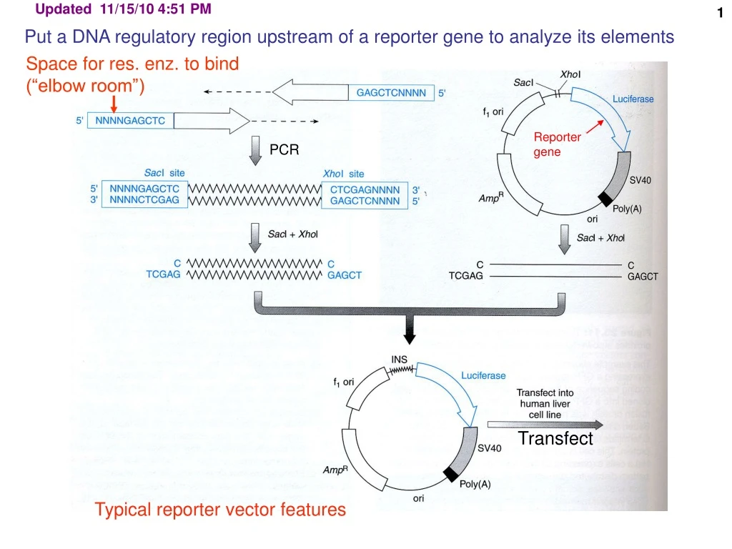
This article provides insights into utilizing DNA regulatory regions upstream of reporter genes for element analysis, including techniques and popular reporters like CAT, Luciferase, and GFP. It covers methodologies such as PCR, transfection, and selective drug resistance for genetic studies.

E N D
Updated 11/15/10 4:51 PM Put a DNA regulatory region upstream of a reporter gene to analyze its elements Space for res. enz. to bind (“elbow room”) Reportergene PCR Transfect Typical reporter vector features
Beta-galactosidase (β-gal) – detection by several different assays Chloramphenicol acetyl transferase (CAT) – detected by a sensitive radioactive assay Luciferase (firefly, Renilla [jellyfish]) – detection, easy dual, sensitive luminescent assay Green fluorescent protein (GFP, BFP, YFP)) – cytological, visible in living cells, fusion proteins, FACS Neomycin phosphotransferase (neo)–selectable drug resistance (geneticin or G418resistance) similarly: resistance to hygromycin, puromycin, histidinol, bleomycin, zeostin Dihydrofolate reductase (DHFR) – selectable in dhfr- cells, amplifiable, fusion proteins work Suicide selection: Herpes simplex virus thymidine kinase (HSVTK) Popular reporters to study promoter/enhancers FACS = fluorescence-activated cell sorter
Gangcyclovir selection AGAINST the presence of enzyme activity (compare to 5-fluoro-orotic acid (FOA) resistance in yeast, URA3-) CRE recombinase (cassette exchange) Mut. protein of interest HSVTK Gancilovir, ATP (non-toxic) Gancilovir-PO4 toxicity, death Mammalian TK Gangcylovir, ATP (Ganciclovir itself is not toxic) Use example: Site-directed recombination Engineered chromosome: lox lox WT protein of interest HSVTK Replacement plasmid: gangcylovir Mut. protein of interest Select recombinants as HSVTK-, ganciclovir-resistant
Testing for a cell-specific promoter: chloramphenicol acetyl transferase (CAT) reporter assay CAT cDNA is from a prokaryotic source. CAT is not found in mammalian cells. Therefore low backgrounds. diacetylated B A Thin layer chromatography (TLC) 14C-chloramphenicol monoacetylated unacetylated Positive control Negative control Applied Molec. Genet., U. Ariz
ONPG (ortho-nitrophenyl-beta-galactoside) – spectrophotometric measurement (420 nm – blue color – simplest) X-gal (5-Bromo-4-chloro-3-indolyl-ß-D-galactoside) – blue precipitate - for cytology or colony detection Umbelliferyl–galactoside (-> umbelliferone, fluorescent, reading in a fluorimeter allows more sensitive quantification than spectrophotometry) Galacton-STAR or some such (-> chemiluminescent product = emission of light, so lower background than fluorescence) Lactose (glucose-beta-galactose disaccharide) – allows growth if hydrolyzed; growth phenotype. For microbial cells usually. Reporter enzyme substrates for different purposes Substrates for beta-galactosidase, for example:
Mapping transcriptional elements upstream of a promoter: Mapping with restrictionenzyme mediated deletions Light units of luciferase in hepatocytes luciferase reporter cDNA gene Conclusion: Applied Molec. Genet., U. Ariz
Footprinting: detects sites on DNA to which protein are bound DNA + DNA-binding protein Naked DNA 32P end-label(e.g., by phosphorylation of the 5’ OH with polynucleotide kinase and gamma 32P-ATP) Population of molecules Partial DNase Population of molecules missing Gel electrophoresis.autoradiography Footprint Many unlabeled fragments are present but not seen
DNA footprint data Note uneven cleavage of naked DNA by DNase Note enhanced cleavage (sensitization) as well as protection
Protein-DNA binding: EMSA or gel shift (EMSA = electrophoretic mobility shift assay) 1 2 3 4 5 competitor (supershift) (shift) DNA element (Even though the hexagon looks like a protein here) Applied Molec. Genet. U. Arizona
Protein-DNA binding: EMSA or gel shift (EMSA = electrophoretic mobility shift assay) http://brc.se.fju.edu.tw/protein/interact/binding.htm
Gel shifts (EMSA (surpershifted complex is not competed by NON-specific probe) Protein DNA complexes migrate more slowly than naked DNA (competed only by specific probe) Super- shift (two molecules of protein bound)
SELEX for protein binding sites on nucleic acids Systematic Evolution of Ligands by Exponential Enrichment Synthesize ~1014 oligomers with a random central section (e.g., 20-mer=1013) ( = 1 microgram =~ several $100) Incubate with a protein of interest Reiterate the cycle 5 to 10 times. If 99.9% efficient: enough Separate the protein-DNA complexes from the free DNA:e.g., using EMSA, IP, nitrocellulose, protein on beads in a column Dissociate complex, PCR amplify the bound DNA fraction Last step: clone and sequence the “winners”
Practical capacity ($700): 1014 random sequences (random ~21-mer = 421) Binding to protein of interest http://www.molmed.uni-luebeck.de/T.%20Restle/Bilder/SELEX.jpg RT
20 nt variable region + two 20 primer templates = 60 nt total length 420 = ~1013 unique sequences 19800 = ~20000 daltons 60 nt X MW of a nucleotide= @330 ug/umole = 0.00005 umoles of the 60-mer X 6.00E+17 molecules per umole molecules = 3E+13 10 ug = 3 x 10¹⁴ molecules, or enough for 30-fold coverage
PUM2, a novel murine puf protein, and its consensus RNA-binding siteWhite EK, Moore-Jarrett T, Ruley HE. RNA. 2001 Dec;7(12):1855-66. Binding site for a “puf “ protein, implicated in mRNA degradation 20-mer Nucleic acid degenerate base abbreviations (FYI) Consensus: Description
TPA = Tissue plasminogen activator, dissolves clots Problem: Cleared quickly from bloodstream by liver Bind to hepatocytes in liver via TPA’s kringle domain Want to isolate a TPA mutant protein with less affinity for hepatocytes Must be still enzymatically active of course.
Goal: to improve tissue plasminogen activator as a therapeutic “clot-busting” treatment Means: Reduce or eiminate the binding of tPA to liver cells, as this clears it from the blood Authors here use a mammalian cells as the carrier of the DNA and the cell surface as a display site. Display was via a fusion protein to a membrane anchor protein, DAF (peptide, really). DAF = “decay accelerating factor” What did they do? Cassette mutagenesis. What region? 333 bp K1 (kringle-1), known to bind the MAb387, which competes for hepatocyte binding (so assuming it is the same target epitope). How did they get kringle mutated? Error-prone PCR How did they isolate just the kringle 1 region? PCR fragment. How did they get the mutagenized fragment back in? Introduced restriction sites at the ends, w/o affecting the coding.
hepatocyte mAb competes with hepatocyte for binding mAb K tPA tPA K K Kringle domain (~100 Aas) tPA
Got this far (two topics through next graphic)
What did they put the mutagenized fragment into? DAF – TPA fusion protein geneHow did they get it into into cells? Electroporation What cells did they use as hosts? 293 carrying SV40 large T antigen How many copies per cell. And why is that important? One, by electroporation at low DNA concentration. [In a transient transfection!] Binding is dominant. Lack of binding (what they are after) is recessive. How did they select cells making MAb387-non-binding TPA? FACS: Recover cells that bind fluorescent mAb vs. protease domainbut low binding to fluorescent mAb vs. kringle domain
Tracked down vector: contains SV40 ori and is transfected into 293 cells making SV40 T-antigen. So plasmid replicates during the transient transfection higher signal.
, Sort the cells with low fluorescence For reiteration of the process
How did they recover the plasmid carrying the mutant TPA gene from the selected cells? Hirt extraction: Like a plasmid prep, lyse cells gently, high MW DNA entangles and forms a “clot”. Centrifuge. Chromosomal DNA soft pellet; plasmid DNA circles stay in supernatant. Then re-transfect, re-sort in FACS. After 2 sorting rounds, test individual E. coli clones: 60% are binding-negative.
MAb to protease domain enriched Collect these No good good good good good Low kringle-1 reactivity MAb to kringle-1 domain FITC = fluorescein reagent. PE = phycoerythrin (fluorescent protein)
Hepatoma cell binding. How? Clone mutated regions into regular TPA gene for testing (no DAF, protein now secreted) Label WT TPA with fluorescein (FITC, conjugated chemically) Mix with hepatoma cells and analyze on a flow cytometer (FACS w/o the sorter part). See specific and non-specific binding. Subtract non-specific binding: the amount not competed by excess un-labeled wt TPA. FITC = fluorescein isothiocyanate
Can’t compete (good) But still haveprotease activity Hepatoma cell binding assay: measure competition for binding of fluorescently labeled WT TPA Binding assay, initial condition No competitor WT Compete. So still bind.