SUBARACHNOID HEMORRHAGE
SUBARACHNOID HEMORRHAGE. www.anaesthesia.co.in anaesthesia.co.in@gmail.com. INTRODUCTION. The presence of blood in the subarachnoid space A syndrome with a number of causes but term usually equated with that occurring secondary to aneurysmal rupture (most common non-traumatic cause).


SUBARACHNOID HEMORRHAGE
E N D
Presentation Transcript
SUBARACHNOID HEMORRHAGE www.anaesthesia.co.inanaesthesia.co.in@gmail.com
INTRODUCTION • The presence of blood in the subarachnoid space • A syndrome with a number of causes but term usually equated with that occurring secondary to aneurysmal rupture (most common non-traumatic cause)
Epidemiology • Overall incidence 10 per 100,000 persons per year • accounts for 3-5% of all strokes • significant cause of morbidity and mortality in previously healthy group of often young people (more potential years life lost than other causes due to earlier mean age of onset, peak 6th decade) • 1.6x higher risk in women • higher incidence in Japan, Finland, and black populations • major risk factors are smoking and potentially hypertension and alcohol abuse
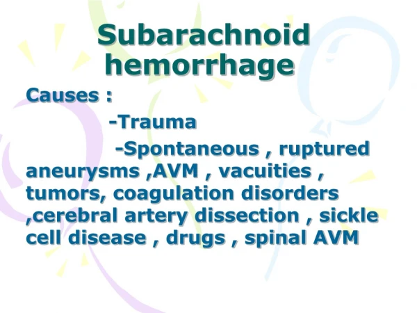
Causes of spontaneous SAH • Ruptured intracranial aneurysms { 75 – 80% } • Cerebral AVMs { 4 – 5% } • Due to tumour • Vasculitides • Cerebral artery dissection { ICA / Vertebral } • Coagulation disorders • Dural sinus thrombosis • SAH of unknown cause { 14 – 22% }
History Headache (HA) • usually a severe ("worst in my life!") HA with onset building to peak severity over seconds (or minutes) • up to half have slower-onset headache building over seconds to few minutes • some have a sentinel bleed with a less persistent but also sudden severe HA in days / weeks preceding ( up to 50% ) • often transient weakness or loss of consciousness at onset (due to abrupt rise in ICP) • others remain obtunded or confused after onset • if predominant neck pain or stabbing between shoulder blades, then suspect SAH due to spinal AVM or fistula
Nausea & Vomiting • often from onset (unlike migraine patient with severe HA but vomiting comes later) • +/- photophobia • +/- neck stiffness • Focal Deficits • stroke-like sudden onset of hemiparesis or other deficit may accompany headache or rarely occurs in its absence
Seizure • 10% or less at onset but can present with this • +/- post-ictal confusional state • if severe post-ictal headache with new-onset seizure, suspect SAH and image • others may not give history of headache if very confused, so need high suspicion
Head Trauma • SAH patients may fall or have an accident as a result of bleeding • this can be confused with traumatic SAH from a trauma itself (eg. Fall ) • hard to establish whether trauma caused the hemorrhage or vice-versa (pattern on CT useful - more at convexity, superficial sulci, adjacent to fracture or impact point) • if no clear cause for accident found (eg. drive off road) then suspect spontaneous SAH first
Ask about • Family History (predisposition found in 5-20%) • Associated disorders include polycystic kidney disease (ADPKD), Ehlers-Danlos IV, Neurofibromatosis type I, +/- Marfan's syndrome (rare causes of aneurysms) • Smoking • Hypertension • Alcohol or illicit drug abuse (e.g. cocaine) • Endocarditis (Hx of fever, malaise in days preceding SAH, known valvular disease) • Hx of head trauma
Examination • Level of consciousness • often depressed, either transiently or usually persisently • important predictor of outcome / severity • use GCS to grade
Pupils / Eyes • monocular blindness can result from large ACom or ophthalmic aneurysm as well as pituitary source of bleeding • assess for oculomotor palsy (pupil fixed & dilated, ptosis, ophthalmoplegia) • can be due to early uncal herniation in comatose patient but more often (in awake SAH patient) due to compression of CN III by aneurysm sac (usually PCom, but also basilar tip and others) • CN VI palsy may be sign of raised ICP (can be bilateral) • look at fundi for papilledema ( NOT seen early ) • the association of vitreous hemorrhage with SAH is known as Terson's syndrome
Focal Motor Deficits • hemiparesis can occur contralateral to side of rupture, usually due to intraparenchymal clot (eg. MCA aneurysm rupturing into Sylvian fissure and peri-insular region) • may also aphasia, neglect, or other deficits • Meningismus: • nuchal rigidity important sign in patient with suspected SAH but takes 6-12 hours to develop • irritation of meninges by blood breakdown products ("chemical meningitis") • may be absent or hard to detect in deeply comatose patients
Systemic Features • may see fever (neurogenic often), hypertensive response (to mainatin cerebral perfusion) and arrhythmias (may contribute to early mortality) as well as almost universal EKG changes
Grading of SAH • WFNS Grading : • GradeGCSMotor Deficit • I 15 Absent • II 13-14 Absent • III 13-14 Present • IV 7-12 +/- • V 3-6 +/-
Serious systemic diseases (hypertension, coronary artery disease, chronic pulmonary disease, diabetes) and severe vasospasm on angiography cause assignment of the patient to the next less favorable category.Hunt WE, Hess EM. J Neurosurg 1968;28:14
Grading System of Fisher 1 No subarachnoid blood detected 2 Diffuse or vertical layers< 1 mm thick 3 Localized clot and/or vertical layer > 1 mm 4 Intracerebral or intraventricular clot with diffuse or no SAH
Predictors of mortality after subarachnoid hemorrhage 1 Poor neurologic condition at hospital admission 2 Depressed level of consciousness after initial bleed 3 Older age4 Preexisting illness5 Elevated blood pressure6 Thick clot in the brain substance or ventricles on initial computed tomographic scan7 Repeat hemorrhage8 Basilar aneurysmal location
Diagnosis • History • Lumbar puncture • CT • MRI • Cerebral angiography • CT angiography ( CTA ) • MR angiography (MRA ) • TCD
Diagnosis • History • Lumbar puncture • CT • MRI • Cerebral angiography • CT angiography ( CTA ) • MR angiography (MRA ) • TCD
Lumbar puncture • Before advent of CT, only modality available • Most sensitive test for SAH • False positive with traumatic tap • Dangerous in patients with raised ICP • the presence of xanthochromia (yellow color) indicates blood breakdown products and cannot be seen in fresh bleeding from traumatic tap (hemoglobin lyses to release OxyHb -> bilirubin)
Diagnosis • History • Lumbar puncture • CT • MRI • Cerebral angiography • CT angiography ( CTA ) • MR angiography (MRA ) • TCD
Nonenhanced CT • First step in the investigation with suspected SAH • Prospective series of 100 patients with suspected SAH 100% within 48 hrs 85% after 5 days 50% after 1 week 30% after 2 weeks • Van Gijn et al. Time course of aneurysmal hemorrhage on CT Neuroradiology 23 : 1985
Contrast enhanced CT -- Helpful in detecting the aetiology of SAH { e.g. aneurysm 30 – 76%, AVM 98 – 100% } • CT – helps in Fishers grading Hydrocephalus infarcts and rebleed
Diagnosis • History • Lumbar puncture • CT • MRI • Cerebral angiography • CT angiography ( CTA ) • MR angiography (MRA ) • TCD
MRI • Not very sensitive in acute stage ( 24 – 48 hrs ) • In subacute and chronic SAH MRI best modality of choice { > 48hrs to over 1 month } • Can detect hemorrhage even after CT and CSF become normal • Contrast enhanced MRI 95 % sensitive { due to formation of methemoglobin which has paramagnetic properties • Also helps in hydrocephalus, mass effects, infarct detection
Diagnosis • History • Lumbar puncture • CT • MRI • Cerebral angiography • CT angiography ( CTA ) • MR angiography (MRA ) • TCD
Cerebral angiography • 4 vessel angio --- gold standard investigation • Helps to identify 1 cause of bleed 2 collateral circulation 3 vasospasm 4 relationship of the cause to parent vessel • Should be done as early as possible after ictus • Can be done under LA / GA
Diagnosis • History • Lumbar puncture • CT • MRI • Cerebral angiography • CT angiography ( CTA ) • MR angiography (MRA ) • TCD
CT angiography • Alternative noninvasive modality to detect etiology of SAH • In aneurysms > 3 mm sensitivity 90 – 95% specificity 100% Harrison MJ et al.neurosurg 40 : 1997
Diagnosis • History • Lumbar puncture • CT • MRI • Cerebral angiography • CT angiography ( CTA ) • MR angiography (MRA ) • TCD
MRA • Probably the best noninvasive modality to detect etiology of SAH • In aneurysms > 3 mm sensitivity 91 – 96% specificity 100% Schwartz RB et al. radiology 192 : 1994 • More useful in patients with angio negative SAH renal failure contrast c/i
Diagnosis • History • Lumbar puncture • CT • MRI • Cerebral angiography • CT angiography ( CTA ) • MR angiography (MRA ) • TCD
Intracranial Aneurysms • Most common cause of SAH • < 2% during childhood • Types 1 saccular { outpouching from arterial bifurcations } 2 fusiform {arterial dilatation } 3 dissecting { pseudoaneurysm } • Ratio of ruptured to unruptured aneurysms 5 : 3 • Etiology : congenital, atherosclerotic, infection, trauma, arteritis etc.
Distribution • occur at vascular branching points • most centered around the circle of Willis (at base of brain, in subarachnoid space) • 20-30% are multiple, and often mirror (eg. bilateral MCA aneurysms) • usually rupture at the dome • Having had an aneurysm rupture (ie. SAH) increases 'risk' of other unruptured aneurysms in same patient rupturing in future
Anterior circulation (85%) • ACA complex incl. Anterior Communicating (AComm) & pericallosal artery • MCA bi-/trifurcation • Internal Carotid Artery (esp at origin of PComm, ophthalmic, anterior choroidal, or terminus) • Posterior Circulation (15%) • mostly at basilar tip • also PICA
Screening for Asymptomatic Aneurysms • may be requested esp in family members of patients with aneursymal SAH or known heritable disorder (esp ADPKD) • strong family history if two or more closely related family members with aneursyms (best to screen first-degree relatives only)
Specific considerations in SAH ICP • Increase rapidly after SAH • May approach SBP • Lasts for several mins and causes LOC • It is thought to limit the amount blood leakage through the ruptured aneurysm • May also increase due to mass effect of hemorrhage or acute hydrocephalus • Correlates well with grade of aneurysm
Impairment of autoregulation and CO2 reactivity • Impaired autoregulation correlates with grade • Impaired autoregulation in presence of vasospasm can lead to DIND • Therefore B P control is very important in SAH pts • CO2 reactivity is generally preserved until there is severe damage • Hyperventilation can be employed perioperatively to reduce CBF and CBV in most of the pts
Intravascular volume and hyponatremia • 36 – 100% pts low intravascular volume • Correlates with grade • Hypovolemia may exacerbate vasospasm and increased DIND • Causes bed rest supine diuresis decreased erythropoiesis iatrogenic blood loss negative nitrogen balance
Hyponatremia • Hyponatremia is seen in 10%–40% of the patients with subarachnoid hemorrhage (SAH) admitted to the neuro critical care unit • Controversy exists regarding both cause and treatment • Hyponatremia is a serum sodium concentration level of less than 135 mEq/L(< 135 mmol/L) for at least 1 day • Less than 120 mEq/L is considered a critical value requiring immediate intervention • Etiology 1 SIADH 2 CSWS
ECG changes • Abnormality in rate and rhythm – 40 to 100% • Usually during first 48 hrs. • Benign to life threatening { VF/VT } • T wave inversion, ST depression, appearance of U waves, prolonged QT interval, Q waves • correlates with amount of blood • Fisher gr 3 & 4 – more ECG changes • Should be differentiated from myocardial ischemia { 32% abnormal thallium scan } • Returns to normal in 10 days { up to 6 wks }
d/d of ECG changes • SAH related benign changes • Myocardial ischemia/ infarction • Electrolyte disturbances hypokalemia hypocalcemia hyponatremia • SAH related hypothalamic injury
Myocardial dysfunction • No correlation with ECG changes • Enzyme studies in case of doubt • Serial Echocardiography After Subarachnoid Hemorrhage • VU Medical Center Academisch Medisch Centrum - Universiteit van Amsterdam. • Study Type: Observational • StudyDesign: Screening,Longitudinal,Defined Population,Prospective Study • Objectives 1) Assessment of the frequency of myocardial dysfunction (segmental wall motion abnormalities, cardiac-specific enzyme elevations, and ECG changes) in patients with SAH. 2) Determination of predictive clinical variables for the occurrence of myocardial dysfunction following SAH. 3) Impact of myocardial dysfunction on neurological prognosis: death, secondary cerebral ischemia, hydrocephalus and rebleeding. • Total Enrollment: 350 Study start: May 2005; Expected completion: January 2009
Respiratory system • Pulmonary edema – due to symapthetic stimulation • Aspiration pneumonia • Others • Systemic hypertension (21%) • Heart disease (3%) • Diabetes mellitus (2%)