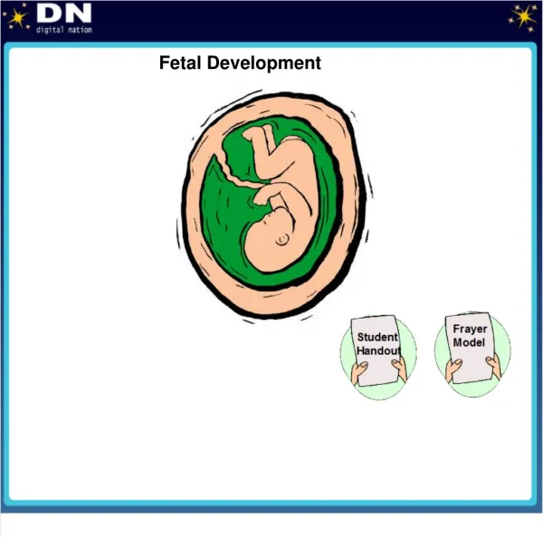
Conception and Fetal Development
Conception and Fetal Development. Cellular Division. Zygote – life begins as a single cell Mitosis – exact copies of the original Meiosis – development of new organism Reproductive cells. Gametogenesis. Oogenesis Process where ovaries form ova

Conception and Fetal Development
E N D
Presentation Transcript
Cellular Division • Zygote – life begins as a single cell • Mitosis – exact copies of the original • Meiosis – development of new organism • Reproductive cells
Gametogenesis • Oogenesis • Process where ovaries form ova • All ova female has is present at 6 months of life • 1st meiotic division secondary oocyte and polar body • At ovulation, 2nd meiotic division begins and completes only if fertilized • One ovum and 3 polar bodies • Spermatogenesis • Process where testes produce sperm • Replicates into two secondary spermatocytes • 2nd meiotic division , 4 spermatids are formed
Egg Sperm Figure 3–1a Gametogenesis involves meiosis within the ovary and testis. During meiosis each oogonium produces a single haploid ovum once some cytoplasm moves into the polar bodies.
Fertilization • Preparation for fertilization • Estrogen levels increase • Peristalsis of fallopian tubes increases • Cervical mucus thins to allow sperm to transfer through • Time frame • Ova viable for 24 hours • Sperm • Capacitation to expose acrosome • Define: Capacitation- Process that removes plasma to expose the acrosome • Define: Acrosome-A membrane at the leading edge of a sperm cell • Block to polyspermy “The penetration of an ovum by more than one sperm” • True fertilization • When nuclei of ovum and sperm unite
Figure 3–2a Sperm penetration of an ovum. The sequential steps of oocyte penetration by a sperm are depicted moving from top to bottom. Source: Scanning electron micrograph from Nilsson, L. (1990). A child is born. New York: Dell Publishing.
Twins • Fraternal • Dizygotic-derived from two separately fertilized eggs • Separate placentas, chorions, amnions (2 of each) • Not identical • Incidence increases with maternal age, in families with genetic factors that increase amounts of gonadotropin • Identical • Monozygotic-derived from a single fertilized egg • Same placenta, chorion and amnion may be the same or different depending on when division occurs • Same sex • Random event
Figure 3–3b Formation of fraternal twins (Note separate placentas.)
Preembryonic development1st 2 wks of development • Cellular Multiplication • Zygote moves through fallopian tube • Rapid mitotic division – morula • Define: Morula- a solid mass of blastomeres that forms when the zygote splits • Blastocyst develops into embryo and amnion • Trophoblast develops into chorion • Implantation (aka “Nidation”- fertilized egg becomes implanted in the lining of the uterus) • Attaches to surface of endometrium (process called,decidua) • Occurs 7 – 9 days after fertilization
Figure 3–4 During ovulation, the ovum leaves the ovary and enters the fallopian tube. Fertilization generally occurs in the outer third of the fallopian tube. Subsequent changes in the fertilized ovum from conception to implantation are depicted.
Embryonic Membranes • Chorion • Outer most membrane • Fingerlike projections, “villi” • Amnion • Thin protective membrane • Contains amniotic fluid • As embryo grows, amnion comes in contact with chorion and forms fluid filled sac
Amniotic Fluid • Functions • Protection, temperature regulation, permit symmetrical growth, prevents adherence of the amnion, gives freedom of movement • Amounts • Ranges from 700 – 1000cc’s at term • Abn Variations: • Oligohydramnios < than normal; Oligo- few, little • Polyhydramnios > 2000cc’s; Poly- many
Preembryonic/Embroyonic • Yolk Sac • Second cavity developed at 8 –9 days • Forms primitive RBC’s during 1st 6 weeks • Umbilical Cord • Formed from the amnion; attaches the embryo to the yolk sac • Three vessels – two arteries and one vein • Wharton’s jelly surrounds vessels in cord preventing cord compression • Twisted or spiraled from fetal movement • Nuchal cord- cord is around the neck • True knot- very rare Lack of Jelly
Figure 3–6 Endoderm differentiates to form the epithelial lining of the digestive and respiratory tracts and associated glands. Source: Adapted from Marieb, E. N. (1998).
PlacentaThere is no mixing between mom’s and baby’s blood • Functions • Metabolic, nutrient, and gas exchange between embryonic and maternal circulation • Development • Chorionic villi – functional layer of placenta • Anchoring villi – forms the partitions/wallscalled cotyledons • Branching villi – vascular system where gas exchange takes place
Placental Circulation • Metabolic gas exchange begins at 4 weeks • Funic souffle soft blowing heard over umbilical cord – synchronous with fetal heartbeat, hearing bloodflow through vessel • Uterine souffle heard just above mother’s pelvis – synchronous with maternal pulse • Placental bloodflow enhanced when mother lies on left side
Placental FunctionsJust know that there’s diff forms of transport • Metabolic activities – produces glycogen, fatty acids are more available, cholesterol, enzymes; stores glycogen, iron • Transport functions • Simple diffusion to • Facilitated transport to requires energy • Active transport to • Pinocytosis transfers large molecules by engulfing • Bulk flow results from hydrostatic and osmotic pressure
Endocrine Functions of the Placenta • Human Chorionic Gonadotropin (HCG) • Prevents normal involution of corpus luteum • Causes corpus luteum to secrete estrogen and progesterone • basis for pregnancy tests • Blood @ 8 –10 days • Urine a few days after missed period
Placental Hormones con. • Progesterone • Hormone of pregnancy • Decreases contractility of the uterus • Maintainance/Essential for pregnancy to continue after 11 weeks • Estrogen • Placenta produces 50% more • Proliferative function – breasts and uterus • Human Placental Lactogen • Similar to pituitary growth hormone • Stimulates changes in maternal metabolic processes • Indicator of fetal growth
(**Will be covered later) Figure 3–11 Fetal circulation. Blood leaves the placenta and enters the fetus through the umbilical vein. After circulating through the fetus, the blood returns to the placenta through the umbilical arteries. The ductus venosus, the foramen ovale, and the ductus arteriosus allow the blood to bypass the fetal liver and lungs.
Embryo and Fetal Development*40wks for pregnancy, 38wks of development • Length of pregnancy • 10 lunar months or 40 weeks (Full term) • Postconception age is 38 weeks after fertilization • Preembryonic • First 14 days of development • Embryonic • Starts on day 15 and continues through 8th week • Most vulnerable to Teratogens
Embryonic StageKNOW: When Hrt Beat? Size? • 3 weeks – broad cephalic end and a narrow caudal end; heart is most advanced organ • 4 – 5 weeks – • vertebrae form from somites; • eyes and ears begin development, arm and leg buds; • partitioning in heart; • brain differentiated into five areas; • heart, brain, and circulatory system show most development
Embryonic Stage con. • 6-7 weeks – head structures highly developed, arms and legs have digits, fetal circulation begins to be established, liver produces RBC’s, eyelids begin to form, rectal and urogenital passages separate • 8 weeks – resembles a human being, eyelids fuse, external genitals appear, rectal passage opens, long bones are forming, and large muscles contract
Figure 3–12 The actual size of a human conceptus from fertilization to the early fetal stage. The embryonic stage begins in the third week after fertilization; the fetal stage begins in the ninth week. Source: Adapted from Marieb, E. N. (1998).
Fetal Stage End of 8th wk • 9-12 weeks – head large and comprises half of fetus, sucking reflex present, limbs are long and slender with well formed digits, forms urine, FHT “Fetal Hrt Tones” heard with doppler, swallows amniotic fluid, meconium present in intestines • 13-16 weeks – rapid growth, lanugo “fine hair”present, moves arms and legs • 20 weeks – weighs about 1 pound, deposits brown fat, head covered with fine wooly hair, eyebrows, eyelids, nails and muscles well developed, mother feels movement quickening
Fetal Stage con. • 24 weeks – weighs 780 gms, hand grasp and startle reflex present, covered with vernix, alveoli in lungs beginning to form • 25-28 weeks – brain develops rapidly, CNS mature enough to provide some regulatory functions, eyelids open, weighs about 1200 gms • 29-32 weeks – pupilary light reflex present, weighs about 2000gms, stores iron, Ca , phosphorous • 36 – 40 weeks – primarily weight gain
Factors Influencing Fetal Development • Quality of ovum or sperm • Adequacy of uterine environment • If unsuitable • Cells may die abortion • Growth slowed • Teratogens “agent/substance which cause malformation”; 1-8wks most vulnerable to induced malformations • Maternal environment • Nutrition– 5th lunar month (20wks) to 6 months of life • Hyperthermia– Sauna/Hot Tub during 1st trimester linked to CNS defects