Human Blastocyst Development and Implantation Process: Week 2
710 likes | 732 Vues
This text provides a detailed overview of the development and implantation process of a human blastocyst during the second week, including the differentiation of trophoblast cells, formation of extraembryonic structures, and establishment of maternal-fetal circulation.

Human Blastocyst Development and Implantation Process: Week 2
E N D
Presentation Transcript
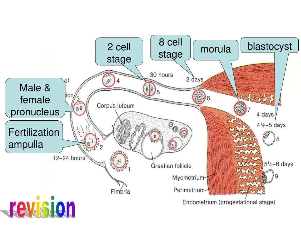
8 cell stage 2 cell stage blastocyst morula Male & female pronucleus Fertilization ampulla revision
Polar trophoblast Zona pellucida inner cell mass blastocoele trophoblast Early blastocyst late blastocyst revision
Day 8 Trophoblast outer layer without distinct cell boundaries, the syncytiotrophoblast inner layer with mononuclear cells, the cytotrophoblast *A 7.5-day human blastocyst, partially embedded in the endometrial stroma.
Day 8 *A 7.5-day human blastocyst *The amniotic cavity appears as a small cleft. Amnioblasts from epiblast (Trophoblast )organize to form a membrane, amnion, which line amniotic cavity. * Endometrial stroma- edematous, highly vascular. Large tortuous glands secrete glycogen and mucus
Bilaminar germ discembryoblast ectoderm (epiblast) Columnar cells endoderm (hypoblast) Cuboidal cells
Day 9 Blastocyst more deeply embedded Fibrin coagulum closes defect in surface epithelium Vacuoles appear in syncitium→fuse → lacunar stage Abembryonic pole flattened cells from hypoblast form thin exocoelomic (Heuser’s)membrane inside cytotrophoblast Hypoblast+ Heuser’s membrane= primitive yolk sac
Day 11 and 12 • Surface epithelium almost entirely covers defect in uterine wall • Lacunar spaces in syncitium- particularly embryonic pole • Synciotrophoblast erode maternal capillaries (sinusoids) • Maternal blood entertrophoblastic system: Uteroplacental circulation
Cells derived yolk sac fill inner surface of cytotrophoblast and outer surface of exocoelomic cavity Fine, loose connective tissue- extraembryonic mesoderm Cavities in extraembryonic mesoderm Cavities confluent→extraembryonic coelom or chorionic cavity Extraembryonic coelom surrounds primitive yolk sac and amniotic cavity except where trophoblast is connected by connecting stalk Day 11 and 12
Day 11 and 12 Extraembryonic somatopleuric mesoderm Lining cytotrophoblast and amnion Extraembryonic splanchnopleuric mesoderm Lining yolk sac
Decidual reaction • decidual response of endometrium • Endometrial changes resulting from adaptation of tissues to implantation of blastocyst • stroma cell → predecidual cell • → decidual cell (cell become polyhedral and rich in glycogen and lipid droplets) • Intercellular spaces filled with extravasate • Tissue edematous
Day 13 • Surface defect closed • Cells of cytotrophoblast proliferate and penetrate syncytiotrophoblast • Cellular column+ syncytial covering= primary villi
Day 13 • Cells from hypoblast migrate inside exocoelomic membrane proloferate and form a new cavity: • secondary yolk sac or definitive yolk sac • Pinched out portions of exocoelomic cavity: exocoelomic cyst
Extraembryonic mesoderm lining inside of cytotrophoblast is chorionic plate. • Extraembryonic mesoderm and two layers of trophoblast form chorion • Chorion forms the wall of chorionic sac in which embryo, its amniotic and yolk sacs are suspended by connecting stalk. Day 13
Day 13 • Extraembryonic mesoderm traverse chorionic cavity in connecting stalk→ development of blood vessels → umbilical cord
By end of 2nd week germ disc : 2 apposed cell discs- • Epiblast: floor of amniotic cavity • Hypoblast: roof of secondary yolk sac Day 13
Day 13 Cephalic region of hypoblastic disc → thickening → columnar cells firmly attached to epiblastic disc → Prochordal plate
Implantation process • zona pellucida disappear (day 5) • Blastocyst attaches to endometrial epithelium (day 6) • Trophoblast differentiates into syncytiotrophoblast and cytotrophoblast (day 7) • Syncytiotrophoblast erodes endometrium and Blastocyst strat embedding (day 8) • Blood filled lacunae appear in syncytiotrophoblast (day 9) • Blastocyst sinks beneath endometrial epithelium (day10) • Lacunar network form by fusion of adjacent lacunae (day 10 & 11) • Syncytiotrophoblast erodes endometrial blood vessels →maternal blood seeps into lacunar network → uteroplacental circulation (day 11 & 12) • Defect in endometrium repaired (day 12 & 13) • Primary chorionic villi develop (day 13 & 14)
Summary of second week • Trophoblast differentiates into syncytiotrophoblast and cytotrophoblast • Decidual reaction: Endometrial changes resulting from adaptation of tissues to implantation of blastocyst • Primary yolk sac forms • Extraembryonic mesoderm forms from cytotrophoblast (yolk sac ) • Extraembryonic coelom • Secondary yolk sac • Amniotic cavity • Bilaminar germ disc: epiblast, hypoblast • Prochordal plate
Second week:The week of twos • Cyto syncytio • Epiblast hypoblast • Somatopleuric splanchnopleuric • Amniotic yolk sac • 2 trophoblast layers • Embryoblast 2 layers • Extraembryonic mesoderm • 2 cavities
Dizygotic twin Diamniotic dichorionic Monozygotic twin (identical twin) -two cell stage: Diamniotic dichorionic -inner cell mass: Diamniotic monochorionic -bilaminar germ disc: Monoamniotic monochorionic Twins
Dizygotic twinMonozygotic twin (identical twin)-two cell stage:Diamniotic dichorionic
Monozygotic twin (identical twin)- inner cell mass:Diamniotic monochorionic
Monozygotic twin (identical twin)- bilaminar germ disc:Monoamniotic monochorionic
Incomplete separation of the inner cell mass gives rise to conjoined twins.
Life man wife
Life papa mom third party intervention
Intrauterine life epiblast man wife hypoblast
Intrauterine life ectoderm papa epiblast mesoderm hypoblast mom endoderm third party intervention
Formation of primitive streak, gastrulation , notochord, Third week of development
Gastrulation: Process by which bilaminar embryonic disc is converted into trilaminar embryonic disc. • Formation of embryonic mesoderm and endoderm. • Begins with formation of primitive streak: thickened linear band on epiblast • Embryo known as gastrula.
primitive streak • Results from proliferation and migration of cells of epiblast to median plane of embryonic disc • Appear:15-16 day • Disappear: 26th day • Cells of both side meet • Elongates by addition of cells from caudal end • Primitive streak helps in identifying craniocaudal axis of embryo
Primitive streak • Cranial end proliferates to form primitive node or Hensen’s node • primitive node - slightly elevated area surrounding small primitive pit • Narrow primitive groove appears on primitive streak continuous with primitive pit (node): invagination of epiblast cells
Electron micrograph showing epiblast cell and primitive node
gastrulation Cells of epiblast migrate towards primitive streak
Gastrulation • On arrival to primitive streak , cells of epiblast become flask- shaped • Detach from epiblast • Slip beneath • invagination
Invaginated cells Lie between epiblast and newly ceated endoderm mesoderm Cells remaining in epiblast ectoderm Displace the hypoblast endoderm
Electron micrograph showing cells of epiblast cells invaginating into hypoblast
Gastrulation Movement of cells beyond limit: laterally cephalad
Invagination of surface cells in primitive streak • Migration forward & laterally continues till end of 4th week • Primitive streak regress ►disappears • Cranial ► differentiation • Caudal ► gastrulation
At the caudal end of the embryo, the primitive streak continues to form mesoderm.
Regions of epiblast migrate and ingress through primitive streak • Cells ingress through cranial region form notochord • Migrating from lateral edge • Cranial end ► paraxial mesoderm • Midstreak ► intermediate mesoderm • Caudal end ►lateral plate mesoderm
Fate of primitive streak • Until 4th week forms intraembryonic mesoderm • Slows down • Diminishes in size • Insignificant structure in sacrococcygeal region • degenerates
Sacrococcygeal teratoma • Remnants of primitive streak persist • Derived from pluripotent primitive streak cells • Elements of 3 germ layers in incomplete stages of differentiation
sirenomelia • Caudal dysgenesis • Insufficient mesoderm in caudalmost part of embryo ??????????????????
Notochord formation • Cellular rod develops by transformation of notochordal process: distinguishable-17-18thday • ► Prenotochordal cells invaginate primitive pit • ► move cephaloid until they reach prechordal plate • ►acquire lumen, notochordal canal
Notochord formation The notochord extends in the midline from the prechordal plate, caudally to the primitive streak