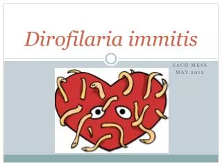
Dirofilaria immitis
By Ryan Hamm and Carolynn Peter. Dirofilaria immitis. Kingdom: Animalia Phylum: Nematoda Class: Secernentea Order: Spirurida Family: Onchocercidae Genus: Dirofilaria Species: immitis. Taxonomy.

Dirofilaria immitis
E N D
Presentation Transcript
By Ryan Hamm and Carolynn Peter Dirofilariaimmitis
Kingdom: Animalia • Phylum: Nematoda • Class: Secernentea • Order: Spirurida • Family: Onchocercidae • Genus: Dirofilaria • Species: immitis Taxonomy
At one time it was confined to the Southern US, it is now found where ever the mosquito vector is found • Worldwide • In US highest infection rates • Along the Gulf and Atlantic coast • Along the Mississippi River GeoGraphic Range
Definitive Hosts: • Canines • Dogs, Foxes, Wolves, Coyotes, • Occasionally cats, rodents, horses, and birds • Accidental Hosts: • Humans • Intermediate Host: • Mosquitoes • Culexpipiens (often infects cats), Anopheles maculipennis, Coquillettidiarichiardii, Aedestriseriatus, Ochlerotatusnotoscriptus (prevalent in Australia), Aedesalbopictus, Aedesaegypti, Culexquinquefasciatus, Aedestaeniorhynchus, Aedesscapularis, Aedestrivittatus Hosts
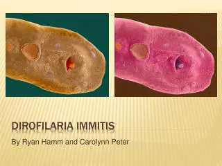
Males • 12-26 cm long • Have a coiled, spiral tail • Females • 25 to 30 cm long • Vulva opening just behind the posterior end of the esophagus • Are ovoviviparous • Microfilariae • 218-329 um long • Have a pointed tail Morphology
Mosquito becomes infected during a blood meal taken from a host containing microfilariae • Microfilarae develop into L3 in the Malpighian tubules and then migrates through the body cavity to the head and mouthparts where they become infective. • The amount of time that this takes depends on temperature • In the mosquito’s next blood meal L3 larvae are deposited into the host. • The molt from L3 to L4 occurs between days 3-12 post infection • L4 molt to final stage occurs at day 50-70 post infection • Worms enter the pulmonary vasculature as early as day 70 and have all arrived by day 90-120. • As they increase in size they occupy larger and larger arteries until they are fully mature • In the right side of the heart, pulmonary arteries, and lungs they mate and produce microfilariae (6-7 months post infection) • The final location of the adult worms depends on the size of the dog and the number of worms present • In humans the worms migrate only to the lungs and forms a lesion. Life cycle
Pathogenesis • On the outside • Loss of appetite • Weight loss • Lethargic • Bulging in the chest • Wheezing and coughing • Vomiting • Diarrhea • Bloody stool • Jaundice • Collapse • On the inside • Large worms extend through the openings of the tricuspid and semilunar valves • Pulmonary arteries show thickening and inflammation of their inner walls • Death occurs from cardiopulmonary failure
Pathogenesis in other Hosts • Cats • only a few worms can be fatal • no treatment • In humans symptoms are unpredictable and vague • Chest pain • Cough • Coughing up blood • Fever • Malaise
Gram negative bacteria • Needed for reproduction and embryogenesis • Pathogenesis of disease • Antibiotic treatment results in sterility of adult worms Wolbachia
Diagnosis • Blood test to detect the female worm antigen • Blood smear to look for microfilariae • ELISA for confirmation • Neither test is consistently positive until 7 months after • X-ray or ultrasound of heart and lungs • Usually done after animal is known to be infected Worms in the heart Heartworm removal
Treatment • Larval worms (L3 and L4) • Ivermectin, milbemycin, selamectin • Must be given every month to prevent the maturation to adult worms • Kills microfilariae if present in infected dog • Suppresses female reproduction • Adults • Melarsominedihydrochloride • Given IM in the lower back • Can be given in 2 or 3 dose sets • Must be done slowly because killing of too many adult worms can cause circulatory shock • Dog can not go for walks or exercise for a month after treatment
Control • Use monthly chemoprophylaxis during mosquito season • Get your dog a yearly heartworm test to ensure that no doses were missed and adult worms have not progressed 4Dx Test
Can result in death for highly infected dogs • High cost of treatment and preventions for pet owners • Has the ability, although rare, to infection humans • Not life threatening, but often confused with lung cancer Public Health Concerns
Quiz What life stages are present in the mosquito? L1, L2, L3 What life stage is infective to the definitive host? L3 Where are infections most prevalent in US? Along the Gulf and Atlantic Coast and the Mississippi What bacteria is needed for heartworm to reproduce? Wolbachia Ivermectin and milbemycin target what life stages? Larval worms (L3 and L4), microfilariae if present In what part of the body do the adult worms live? Right side of the heart and right pulmonary arteries
http://plpnemweb.ucdavis.edu/nemaplex/Taxadata/Dimmitis.htm • http://plpnemweb.ucdavis.edu/nemaplex/Taxadata/Dimmitis.htm • http://www.marvistavet.com/html/body_heartworm_treatment.html • http://www.wolbachia.sols.uq.edu.au/about.cfm • http://www.heartwormsociety.org/ • Foundations of Parasitology by Gerald Schmidt and Larry S. Roberts Sources