Download
1 / 18
180 likes | 444 Vues
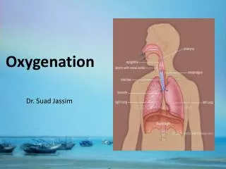
Case Discussion- Oxygenation Conundrums. Dr. TH de Klerk. Goals of Ventilation. Oxygenation: PaO2 >60mmHG, Sats >90%.

E N D
Case Discussion- Oxygenation Conundrums Dr. TH de Klerk
Goals of Ventilation • Oxygenation: PaO2 >60mmHG, Sats >90%. • Lung protective ventilation: Tidal volume< 6ml/kg IBW (male height in cm- 100, female height in cm- 110). Plateau pressure < 30cm H2O. Transpulmonary plateau pressure<25cm H20. PEEP set acc to best dynamic compliance- not >15cm H2O, except when using PEEP set acc to transpulmonary expiratory pressure +2-3cm H2O. • Reverse cause for respiratory failure. • Patient comfort, decrease work of breathing. Avoid dyssynchrony (trigger, flow, cycling). • Acid- Base status: PCO2 < 120mmHg, pH >7.20
Case Study 1 • Ms. MS, 33yr African female. Background HIV positive on HAART (TDF, 3TC, EFV) since 2009. CD4: 298 with undetectable viral load- Dec 2013. History of Pulmonary Tuberculosis 2010- treated for 6months. Secondary bronchiectasis with pulmonary hypertension. Currently 22wks pregnant. • Now multi-lobar, community acquired pneumonia, CURB 65 score: 3, intubated and ventilated for a mixed Type 1 and 2 respiratory failure. Clinical features of right heart failure. • CT Pulmonary angiogram couldn’t demonstrate pulmonary embolus, infiltrates suggestive of atypical pneumonia. Patient also treated for Pneumocystis pneumonia. • Echocardiography demonstrated dilatation and systolic dysfunction of right ventricle.
Case study 1- cont. • Patients clinical picture further complicated by acute pancreatitis, CT suggestive of pancreatic oedema, no necrosis. Intra- abdominal pressure 13mmHg (18cm H20). • Oxygenation worsened: P/F ratio of 60, PEEP: 10cm H20, with Peak Pressure of 35cm H20. Plateau pressure 28cm H2O • Unfortunately no oesophageal probes available at that time. • PEEP was increased to 20cm H20, Peak Pressure at 40cm H20. Plateau pressure: 35cm H2O. Oxygenation indices immediately improved, P/F ratio went up to 150.
Case Study 1- Discussion • What is the goals of ventilation in a pregnant patient with ARDS? • What should we aim for in ventilating patients with pulmonary hypertension? • What should we aim for in ventilating patients with intra-abdominal hypertension?
ARDS in Pregnancy • 40% maternal mortality rate, 25% perinatal fetal mortality rate. • Certain causes unique to pregnancy. • Pregnancy altered physiology e.g. respiratory drive, FRC, etc. • Definition: ARDS occurring during pregnancy, results from an obstetric cause or is otherwise modified by an obstetric-related factor.
ARDS in Pregnancy Commonest causes: • Sepsis (most common): pyelonephritis, aspiration, chorio-amnionitis. • Pre-eclampsia. • Amniotic fluid embolism. • Obstetric related hemorrhage: abruptio, etc. • Tocolytic- induced pulmonary edema.
Ventilation in Pregnancy • Goals of ventilation differ! • PaO2> 70mmHg, Sats > 95%. • PaCO2< 45mmHg. Needs a gradient between mother and baby. • 7.45< pH< 7.30 • Placental insufficiency- vasoconstriction placental bed. • intra- abdominal pressure with decreased chest wall compliance (vascularity and edema)- tolerate higher inspiratory plateau pressure. • Prone positioning- not feasible.
Ventilation in Pulmonary hypertension • ARDS is most common cause for new onset pulmonary hypertension in ICU. • The reason for decreased mortality in ARDS due to lung protective ventilation thought to be due to decreased incidence acute corpulmonale. • Prevent hypoxemia- esp. low mixed venous O2 saturation. • Aim for PCO2< 55mmHg. • Avoid pH< 7.25 • Treat sepsis- causes pulmonary vasoconstriction. • Lung protective ventilation- tidal volume <6ml/kg IBW, Pplat <27cm H2O, PEEP optimize to best dynamic compliance. PEEP and Mean airway pressure- compress pulmonary capillaries. • Treat intra-abdominal hypertension.
Ventilation Intra-abdominal hypertension. • 25-80% of IAP transmitted to intrathoracic compartment- not predictable. • Mechanical ventilation intra-abdominal pressure 1-2mmHg. Increasing PEEP add additional 1-2mmHg intra- abdominal pressure. • chest wall elastance, intra-pleural pressure= transpulmonary pressure. Recommend using esophageal pressure. • Prone position compression of abdomen. • Elevating head 45°-IAP 5-15mmHg. • Reverse Trendelenburg position- improve lung mechanics, but splachnic perfusion. • Setting PEEP in cmH2O= IAP in mmHg. Or setting PEEP @ ½ IAP in cmH20. Only rough guide.
Case Study 2 • Mr. JDB, 38yr Caucasian male. Background VSD repair at age 20 (not palliative),morbid obesity (BMI:68), proportional short stature (5ft, 3inch), poorly controlled hypertension, dyslipidemia and impaired glucose tolerance. • Presented acute pulmonary edema with mixed type 1and 2 respiratory failure requiring intubation and ventilation. • Collateral history from wife suggestive of obstructive sleep apnea- excessive day time somnolence, nocturnal apneic episodes.
Case Study 2- cont • Patients optimal PEEP determined with trans-pulmonary pressure measurement: 16cm H20. • Echocardiogram limited: LVEF:40%, no significant residual VSD and possible pulmonary hypertension. • Oxygenation indices improved over next few days, but PCO2 remained 55-70mmHg. Patient awake and calm, no increased respiratory rate or increased work of breathing. Possible central sleep apnea. • Patient subsequently weaned and extubated.
Case Study 2- Discussion • How to determine the optimal PEEP for patient with morbid obesity? • During the weaning phase, what is the optimal level of PEEP?
Ventilation in Obesity • BMI ∞ inflammatory mediators- esp. bronchospasm. • ARDS H1N1 mortality- X 5-15. • TLC, FRC, VC, pleural pressure,upper and lower airway resistance, intra-abdominal pressure. • Hypoxemia- Atelectasis and V/Q mismatch due to airway narrowing. • Pplat <30cm H2O not adequate- due to pleural pressure. Pplat poor surrogate for transpulmonary pressure in this setting. Use esophageal pressure measurements to set PEEP and transpulmonary pressure. • Some studies recommend ‘empiric’ PEEP of 10-14. • Ventilate in sitting position. • NB cause dynamic hyperinflation- set PEEP to 80% of Auto-PEEP. Counter act collapsed airway.
Ventilation in CNS injury • Neurogenic pulmonary edema- sympathetic storm with myocardial stunning. • Hyperventilation global brain oxygenation-not good idea ischaemic stroke, cardiac arrest. • Hypoventilation cerebral blood flow- hemorrhage in patient with intracranial bleeding. • Diffuse cerebral edema- hyperventilation to PCO2= 30-35mmHg used if imminent herniation- temporary effect. • PEEP+ MAP= ICP, not linear effect. Due to decreased venous return. • Avoid bronchoscopy in patients with ICP. • Don’t extubate high spinal cord injuries within first 72hrs- cord edema, extending injury.
References • Hibbert K, Rice M, Malhotra A. Obesity and ARDS. Chest 2012 Sep; 142(3): 785-790. • Cole DE, Taylor TL, McCullough DM, Shoff CT, Derdak S. Acute Respiratory Distress Syndrome in pregnancy. Crit Care Med 2005 Oct; 33 (10 Suppl): S269-278. • Esquinas AM, Petroianni A. Pulmonary hypertension in critically ill patients with mechanical ventilation: still a greatest challenge for intensivists. J Crit Care 2014 Feb: 29(1): 166. • Stevens RD, Lazaridis C, Chaleta JA. The role of mechanical ventilation in acute brain injury.NeurolClin 2008 May; 26(2):543-563. • Malbrain M, De Waele J. Intra- abdominal Hypertension. Core Critical Care Series 2013.