Download
1 / 58
580 likes | 665 Vues
Learn about the importance of distinguishing TIAs, risk factors, and differential diagnosis. Understand the presentation, symptoms related to cerebral circulation, and other conditions to consider. This guide provides valuable insights for healthcare professionals.

E N D
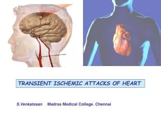
Transient Ischemic Attacks Rodney W. Smith, MDClinical Assistant ProfessorDepartment of Emergency MedicineUniversity of MichiganAnn Arbor, MI
Example Case • A 55 year old male presents to the emergency department with acute onset of • Left arm weakness: Unable to lift left arm off of lap • Symptoms improved on the way to the hospital
Example Case • PMHx: Hypertension • Takes enalapril • ROS: • No headache • No other neurologic symptoms • Social Hx: • Smokes 1 ppd
Example Case • Physical Exam • Overweight, in NAD • 160/90, 80, 14, 37.5C • Right carotid bruit • Heart with regular rate and rhythm; No murmur
Example Case • Neuro exam • Oriented to person, place, and time • Fluent speech • CN II-XII intact • Motor 4/5 strength in left upper extremity • Sensory subjective decrease in pinprick in left upper extremity compared to the right • DTR +2 except at left biceps +3 • Gait steady • Cerebellar intact finger to finger and finger to nose • No extensor plantar response.
Summary • Importance of distinguishing TIA from other causes of transient “spells” • Essential elements include a careful history, physical exam, and CT scan • ED treatment and disposition are directed toward prevention of subsequent stroke • Incidence of early stroke after TIA justifies hospital admission for further evaluation
Risk Factors/Epidemiology • 300,000 TIAs per year in US • 5-year stroke risk after TIA 29% • 43.5% in 2 years with >70% carotid stenosis treated medically • Many stroke patients have had TIA • 25% - 50% in large artery atherothrombotic strokes • 11% - 30% in cardioembolic strokes • 11% to 14% in lacunar strokes
Risk Factors/Epidemiology • Risk factors are the same as stroke • Increasing age • Sex • Family history / Race • Prior stroke / TIA • Hypertension • Diabetes • Heart disease • Carotid artery / Peripheral artery disease • Obesity • High cholesterol • Physical inactivity
ED Presentation • What is a TIA? • Acute loss of focal cerebral function • Symptoms last less than 24 hours • Due to inadequate blood supply • Thrombosis • Embolism
ED Presentation • Acute loss of focal cerebral function • Motor symptoms • Weakness or clumsiness on one side • Difficulty swallowing • Speech disturbances • Understanding or expressing spoken language • Reading or writing • Slurred speech • Calculations
ED Presentation • Acute loss of focal cerebral function • Sensory symptoms • Altered feeling on one side • Loss of vision on one side • Loss of vision in left or right visual field • Bilateral blindness • Double vision • Vertigo
ED Presentation • Non-focal Symptoms (Not TIA) • Generalized weakness or numbness • Faintness or syncope • Incontinence • Isolated symptoms (symptoms occurring alone) • Vertigo or loss of balance • Slurred speech or difficulty swallowing • Double vision
ED Presentation • Non-focal Symptoms (Not TIA) • Confusion • Disorientation • Impaired attention/concentration • Diminution of all mental activity • Distinguish from • Isolated language or visual-spatial perception problems (may be TIA) • Isolated memory problems (transient global amnesia)
ED Presentation • Acute loss of focal cerebral function • Abrupt onset • Symptoms occur in all affected areas at the same time • Symptoms resolve gradually • Symptoms are “negative”
ED Presentation • Symptoms last less than 24 hours • Most last less than one hour • Less than 10 percent > 6 hours • Amaurosis fugax up to five minutes
ED PresentationDifferential Diagnosis • Migraine with aura • Positive symptoms • Spread over minutes • Visual disturbances • Somatosensory or motor disturbance • Headache within 1 hour
ED PresentationDifferential Diagnosis • Aura without Headache • 98% Visual symptoms • 30% with other symptoms • 26% sensory • 16% aphasia • 6% dysarthria • 10% weakness • Mean age 48.7 (vs. 62.1) • Fewer cardiovascular risk factors
ED PresentationDifferential Diagnosis • Partial (focal) seizure • Positive sensory or motor symptoms • Spread quickly (60 seconds) • Negative symptoms afterward (Todd’s paresis) • Multiple attacks
ED PresentationDifferential Diagnosis • Transient global amnesia • Sudden disorder of memory • Antegrade and often retrograde • Recurrence 3% per year • Etiology unclear • Migraine • Epilepsy (7% within 1 year) • Unknown
ED PresentationDifferential Diagnosis • Transient global amnesia • No difference in vascular risk factors compared with general population • Fewer risk factors when compared with TIA patients • Prognosis significantly better than TIA
ED PresentationDifferential Diagnosis • Structural intracranial lesion • Tumor • Partial seizures • Vascular steal • Hemorrhage • Vessel compression by tumor
ED PresentationDifferential Diagnosis • Intracranial hemorrhage • ICH rare to confuse with TIA • Subdural hematoma • Headache • Fluctuation of symptoms • Mental status changes
ED PresentationDifferential Diagnosis • Multiple sclerosis • Usually subacute but can be acute • Optic neuritis • Limb ataxia • Age and risk factors • Signs more pronounced than symptoms
ED PresentationDifferential Diagnosis • Labyrinthine disorders • Central vs. Peripheral vertigo • Ménière's disease • Benign positional vertigo • Acute vestibular neuronitis
ED PresentationDifferential Diagnosis • Metabolic • Hypoglycemia • Hyponatremia • Hypercalcemia • Peripheral nerve lesions • Entrapments • Painful quality
ED PresentationDifferential Diagnosis • Patient evaluation by senior neurologists with interest in stroke • Agreement on 48 of 56 patients (85.7%) • 36 with TIA • 12 Not TIA • 8 of 56 disagreement • 4 of these, both listed firm diagnosis
ED Diagnosis and Evaluation • History • Characteristics of the attack • Associated symptoms • Risk factors • Vascular Disease • Cardiac Disease • Hematologic Disorders • Smoking • Prior TIA
ED Diagnosis and Evaluation • Physical Examination • Neurologic Exam • Carotid Bruits • Cardiac Exam • Peripheral Pulses
ED Diagnosis and Evaluation • EKG • CBC, Coags, and Chemistries • Chest Xray • Head CT without contrast • Expedite if early presentation
ED Diagnosis and Evaluation • Symptom vs. Disease • Significant carotid artery stenosis • Cardiac embolism • Admission vs. Discharge • Traditional approach • Trend toward outpatient evaluation
ED Diagnosis and Evaluation • Stroke Rate After TIA • Percent (95% CI)
ED Diagnosis and Evaluation • Stroke Rate After TIA • Johnston, et al. JAMA 284:2901, 2000. • Follow-up of 1707 ED patients diagnosed with TIA • Stroke rate at 90 days was 10.5% • Half of these occurred in the first 48 hours after ED presentation
Management • Goal: Prevention of Stroke • Expedited Evaluation • Carotid Artery Disease • Cardioembolism • Inpatient vs. Observation Unit vs. Outpatient • Antiplatelet Therapy • Risk Factor Modulation
ManagementED Disposition • Discharge • Further testing will not change treatment • Prior workup • Not a candidate for CEA or anticoagulation
ManagementED Disposition • Admission • Clear indication for anticoagulation • Severe deficit • Crescendo symptoms • Other indication for admission • Admission or observation unit evaluation • All others
ManagementDiagnosis of Carotid Stenosis • Carotid Duplex Ultrasound • Sensitivity of 94 - 100% for > 50% stenosis • May overdiagnose occlusion • Non-invasive
ManagementDiagnosis of Carotid Stenosis • Magnetic Resonance Angiography • Similar sensitivity to carotid ultrasound • Overestimates degree of stenosis • Gives information about vertebrobasilar system • Accuracy of 62% in detecting intracranial pathology • Cost and claustrophobia
ManagementDiagnosis of Carotid Stenosis • Cerebral Angiography • Gold standard for diagnosis • Invasive, with risk of stroke of up to 1% • For patients with positive ultrasound • For patients with occlusion on ultrasound • First test if intracranial pathology suspected
ManagementCardiogenic Embolism • Major risk factors: Anticoagulation Indicated • Atrial fibrillation • Mitral stenosis • Prosthetic cardiac valve • Recent MI • Thrombus in LV or LA appendage • Atrial myxoma • Infective endocarditis (No anticoagulation) • Dilated cardiomyopathy
ManagementCardiogenic Embolism • Minor risk factors: Best treatment unclear • Mitral valve prolapse • Mitral annular calcification • Patent foramen ovale • Atrial septal aneurysm • Calcific aortic stenosis • LV regional wall motion abnormality • Aortic arch atheromatous plaques • Spontaneous echocardiographic contrast
ManagementEchocardiogram • Yield < 3% in undifferentiated patients • Higher with risk factors • TEE preferred • Specific treatment of many abnormalities unknown
ManagementEchocardiogram • Indications • Age < 50 • Multiple TIAs in more than one arterial distribution • Clinical, ECG, or CXR evidence suggests cardiac embolization
Management TIA with Atrial Fibrillation • INR 2.5 (Range 2 to 3) • Aspirin if Warfarin contraindicated • Timing of onset of AC not proven in RCT • AC in other causes of cardioembolic stroke not proven in RCT EAFT Study Group, Lancet, 1993
ManagementAntiplatelet Therapy • Aspirin • Compared with placebo in patients with minor stroke/TIA • Relative risk of composite endpoint reduced by 13% to 17% • Dose of aspirin probably not important • Lower dose gives lower incidence of GI side effects.
Management • Ticlopidine • Small absolute risk reduction compared with ASA • Side effects preclude use in up to 5% • Serious adverse effects • Neurtropenia • Thrombotic thrombocytopenic purpura
Management • Clopidogrel • Similar to Ticlopidine in reducing composite endpoint • Reduction in risk of stroke alone less than with Ticlopidine • Similar side effect profile to ASA