Download
1 / 22
240 likes | 767 Vues
The extraembryonic mesoderm lining the inside of the cytotrophoblast is then known as THE CHORIONIC PLATE The only place where extraembryonic mesoderm traverses the chorionic cavity is in THE CONNECTING STALK With development of blood vessels, the stalk becomes THE UMBILICAL CORD.

E N D
The extraembryonic mesoderm lining the inside of the cytotrophoblast is then known as THE CHORIONIC PLATE The only place where extraembryonic mesoderm traverses the chorionic cavity is in THE CONNECTING STALK With development of blood vessels, the stalk becomes THE UMBILICAL CORD
THIRD WEEK OF DEVELOPMENT
It is not birth, marriage or death, but gastrulation, which is truly the most important time in your life Lewis Wolpert (1986)
When viewed from above, through the amniotic cavity, the epiblast appears as an oval disc
The BUCCOPHARYNGEALMEMBRANE marks the future mouth which is situated in the midline at the cranial end The CLOACAL MEMBRANE marks the future anus which is situated in the midline at caudal end.
The cells of the EPIBLAST are capable of proliferation and migration These two features of the epiblast will lead to: • The cells of the epiblast start to proliferate forming • a swilling called PRIMITIVE NODE
As the primitive node elongates THE PRIMITIVE STREAK appears • Cells of the epiblast migrate toward the primitive streak . • The cells of the primitive streak ingress in the epiblast making a pore in the middle • Upon arrival in the region of the streak, they detach from the epiblast, and slip beneath it. • This inward movement is known as invagination.
Once the cells have invaginated, some displace the hypoblast, creating the embryonic ENDODERM • Other cells come to lie between the epiblast and newly created endoderm to form MESODERM • Cells remaining in the epiblast then form ECTODERM. • Thus, THE EPIBLAST, through the process of gastrulation, • is the source of all of the germ layers. • cells in these layers will give rise to all of the tissues and organs in the embryo.
A swelling appears on the upper surface of the hypoblast called NOTOCHORD Because of the presence of the notochord in the middle of the trilaminar disc , the migrating cells from the epiblast will fill only the paraxial region (the area around the axis)
The most characteristic event occurring during the third week of gestation is GASTRULATION, the process that establishes all three germ layers in the embryo ENDODERM 1-ECTODERM 2-MESODERM 3-ENDODERM
Derivatives of the ectodermal germ layer
Development of the neural tube At the middle of the epiblast another swelling called 1- neural plate appears The neural plate replaces the receding primitive streak and closes the pore formed before
Parts of the neural tube 1-neural crest 2-alar plate 3-basal plate alar plate basal plate
The nervous system is formed from the ectoderm (the neural tube) The neural crest gives rise to the ganglia The alar plate gives rise to the sensorypart of the nervous system The basal plate gives rise to the motor part of the nervous system
Neural Crest Derivatives 1-Connective tissue and bones of the face and skull 2-Cranial nerve ganglia 3-C cells of the thyroid gland 4-Conotruncal septum in the heart 5-Odontoblasts 6-Dermis in face and neck 7-Spinal (dorsal root) ganglia 8-Sympathetic chain and preaortic ganglia 9-Parasympathetic ganglia of the gastrointestinal tract 10-Adrenal medulla 11-Schwann cells 12-Glial cells 13-Arachnoid and pia mater (leptomeninges) 14-Melanocytes
The notochord gives rise to the Nucleus pulpous Of the intervertebral disk
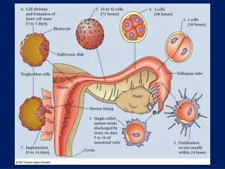
MAJOR EVENTS OF THE SECOND WEEK OF DEVELOPMENT • A-Completion of implantation of the blastocyst • B-Production of a bilaminar embryonic disc • C-FORMATION OF EXTRAEMBRYONIC STRUCTURES: • 1-AMNIOTIC CAVITY • 2-AMNION • 3-YOLK SAC • 4-CHORIONIC SAC • 5- CONNECTING STALK • D-APPEARANCE OF PRIMARY CHORIONIC VILLI • E-APPEARANCE OF THE PRECHORDAL PLATE
MAJOR EVENTS OF THE THIRD WEEK • Development of THE PRIMITIVE STREAK • Development of THE NOTOCHORD • Formation of THE TRILAMINAR GERM DISC • Beginning of formation OF NEURAL TUBE