Download
1 / 162
1.64k likes | 2.11k Vues
17 The Special Senses. An Introduction to the Special Senses. Learning Outcomes 17-1 Describe the sensory organs of smell, trace the olfactory pathways to their destinations in the brain, and explain the physiological basis of olfactory discrimination.

E N D
17 The Special Senses
An Introduction to the Special Senses • Learning Outcomes • 17-1 Describe the sensory organs of smell, trace the olfactory pathways to their destinations in the brain, and explain the physiological basis of olfactory discrimination. • 17-2 Describe the sensory organs of taste, trace the gustatory pathways to their destinations in the brain, and explain the physiological basis of gustatory discrimination. • 17-3 Identify the internal and accessory structures of the eye, and explain the functions of each.
An Introduction to the Special Senses • Learning Outcomes • 17-4 Explain color and depth perception, describe how light stimulates the production of nerve impulses, and trace the visual pathways to their destinations in the brain. • 17-5 Describe the structures of the external, middle, and internal ear, explain their roles in equilibrium and hearing, and trace the pathways for equilibrium and hearing to their destinations in the brain.
An Introduction to the Special Senses • Five Special Senses • Olfaction • Gustation • Vision • Equilibrium • Hearing
17-1 Smell (Olfaction) • Olfactory Organs • Provide sense of smell • Located in nasal cavity on either side of nasal septum • Made up of two layers • Olfactory epithelium • Lamina propria
17-1 Smell (Olfaction) • Layers of Olfactory Organs • Olfactory epithelium contains: • Olfactory receptors • Supporting cells • Basal (stem) cells
17-1 Smell (Olfaction) • Layers of Olfactory Organs • Lamina propria contains: • Areolar tissue • Blood vessels • Nerves • Olfactory glands
Figure 17-1a The Olfactory Organs Olfactory Pathway to the Cerebrum Olfactoryepithelium Olfactorynervefibers (N I) Olfactorytract Centralnervoussystem Olfactorybulb Cribriformplate Superiornasalconcha The olfactory organ onthe left side of the nasal septum
Figure 17-1b The Olfactory Organs Basal cell:divides to replaceworn-out olfactoryreceptor cells Toolfactorybulb Olfactorygland Cribriformplate Olfactorynerve fibers Laminapropria Developingolfactoryreceptor cell Olfactoryreceptor cell Olfactoryepithelium Supporting cell Mucous layer Knob Olfactory cilia: surfaces containreceptor proteins(see SpotlightFig. 173) Subsance being smelled An olfactory receptor is a modifiedneuron with multiple cilia extendingfrom its free surface.
17-1 Smell (Olfaction) • Olfactory Glands • Secretions coat surfaces of olfactory organs • Olfactory Receptors • Highly modified neurons • Olfactory reception • Involves detecting dissolved chemicals as they interact with odorant-binding proteins
17-1 Smell (Olfaction) • Olfactory Pathways • Axons leaving olfactory epithelium • Collect into 20 or more bundles • Penetrate cribriform plate of ethmoid • Reach olfactory bulbs of cerebrum where first synapse occurs
17-1 Smell (Olfaction) • Olfactory Pathways • Axons leaving olfactory bulb: • Travel along olfactory tract to reach olfactory cortex, hypothalamus, and portions of limbic system • Arriving information reaches information centers without first synapsing in thalamus
17-1 Smell (Olfaction) • Olfactory Discrimination • Can distinguish thousands of chemical stimuli • CNS interprets smells by the pattern of receptor activity • Olfactory Receptor Population • Considerable turnover • Number of olfactory receptors declines with age
Figure 17-2 Olfactory and Gustatory Receptors Olfaction and gustation are specialsenses that provide us with vitalinformation about our environment. Although the sensory information provided is diverse and complex, each special sense originates at receptor cells that may be neurons or specialized receptor cells that communicate with sensory neurons. Stimulus Dendrites Specializedolfactoryneuron Stimulusremoved Actionpotentials Stimulus Threshold Generator potential to CNS
Figure 17-2 Olfactory and Gustatory Receptors Olfactory reception occurs on the surface membranes ofthe olfactory cilia. Odorantsdissolved chemicals thatstimulate olfactory receptorsinteract with receptors calledodorant- binding proteins on the membrane surface. In general, odorants are small organic molecules. Thestrongest smells are associated with molecules of eitherhigh water or high lipid solubilities. As few as fourodorant molecules can activate an olfactory receptor. The binding of an odorant to itsreceptor protein leads to theactivation of adenylyl cyclase, theenzyme that converts ATP tocyclic-AMP (cAMP). The cAMP then openssodium channels in theplasma membrane, which,as a result, begins todepolarize. If sufficient depolarizationoccurs, an action potential istriggered in the axon, and theinformation is relayed to theCNS. MUCOUSLAYER Odorantmolecule Closedsodiumchannel Depolarizedmembrane Activeenzyme Inactiveenzyme Sodiumions enter RECEPTORCELL
17-2 Taste (Gustation) • Gustation • Provides information about the foods and liquids consumed • Taste Receptors (Gustatory Receptors) • Are distributed on tongue and portions of pharynx and larynx • Clustered into taste buds
17-2 Taste (Gustation) • Taste Buds • Associated with epithelial projections (lingual papillae) on superior surface of tongue
17-2 Taste (Gustation) • Three Types of Lingual Papillae • Filiform papillae • Provide friction • Do not contain taste buds • Fungiform papillae • Contain five taste buds each • Circumvallate papillae • Contain 100 taste buds each
17-2 Taste (Gustation) • Taste Buds • Contain: • Basal cells • Gustatory cells • Extend taste hairs through taste pore • Survive only 10 days before replacement • Monitored by cranial nerves that synapse within solitary nucleus of medulla oblongata • Then on to thalamus and primary sensory cortex
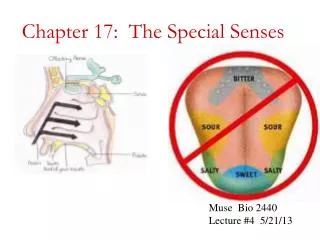
Figure 17-3a Gustatory Receptors Water receptors(pharynx) Umami Sour Bitter Salty Sweet Landmarks andreceptors on thetongue
Figure 17-3b Gustatory Receptors Tastebuds Circumvallate papilla Fungiform papilla Filiform papillae The structure and representative locationsof the three types of lingual papillae. Tastereceptors are located in taste buds, whichform pockets in the epithelium of fungiform or circumvillate papillae.
Figure 17-3c Gustatory Receptors Tastebuds Taste buds LM 280 Nucleus oftransitional cell Nucleus ofgustatory cell Nucleus ofbasal cell Taste bud LM 650 Transitional cell Gustatory cell Taste hairs(microvilli) Basal cell Taste pore Taste buds in a circumvallate papilla.A diagrammatic view of a taste bud,showing gustatory (receptor) cellsand supporting cells.
17-2 Taste (Gustation) • Gustatory Discrimination • Four primary taste sensations • Sweet • Salty • Sour • Bitter
17-2 Taste (Gustation) • Additional Human Taste Sensations • Umami • Characteristic of beef/chicken broths and Parmesan cheese • Receptors sensitive to amino acids, small peptides, and nucleotides • Water • Detected by water receptors in the pharynx
17-2 Taste (Gustation) • Gustatory Discrimination • Dissolved chemicals contact taste hairs • Bind to receptor proteins of gustatory cell • Salt and sour receptors • Chemically gated ion channels • Stimulation produces depolarization of cell • Sweet, bitter, and umami stimuli • G proteins • Gustducins
17-2 Taste (Gustation) • End Result of Taste Receptor Stimulation • Release of neurotransmitters by receptor cell • Dendrites of sensory afferents wrapped by receptor membrane • Neurotransmitters generate action potentials in afferent fiber
17-2 Taste (Gustation) • Taste Sensitivity • Exhibits significant individual differences • Some conditions are inherited • For example, phenylthiocarbamide (PTC) • 70% of Caucasians taste it but 30% do not • Number of taste buds • Begins declining rapidly by age 50
Figure 17-2 Olfactory and Gustatory Receptors Receptor cell Stimulus Stimulusremoved Stimulus Receptorcell Threshold Receptor depolarization Synapse Axon ofsensoryneuron Axon Actionpotentials Stimulus Synapticdelay to CNS Generator potential
Figure 17-2 Olfactory and Gustatory Receptors Sweet, Bitter, and Umami Receptors Salt and Sour Receptors Receptors responding to stimuli that produce sweet, bitter, and umami sensations are linked to G proteins called gustducins (GUST-doos- inz)protein complexes that use second messengers to produce their effects. Salt receptors and sour receptors are chemically gated ion channels whose stimulation produces depolarization of the cell. Sweet,bitter, orumami Sour,salt Membranereceptor Gated ionchannel Resting plasmamembrane InactiveG protein ActiveG protein Channel opens Depolarizedmembrane ActiveG protein Active2nd messenger Inactive2nd messenger Activation of second messengers stimulates release of chemical neurotransmitters. Depolarization of membranestimulates release of chemicalneurotransmitters.
17-3 Accessory Structures of the Eye • Accessory Structures of the Eye • Provide protection, lubrication, and support • Include: • The palpebrae (eyelids) • The superficial epithelium of eye • The lacrimal apparatus
17-3 Accessory Structures of the Eye • Eyelids (Palpebrae) • Continuation of skin • Blinking keeps surface of eye lubricated, free of dust and debris • Palpebral fissure • Gap that separates free margins of upper and lower eyelids
17-3 Accessory Structures of the Eye • Eyelids (Palpebrae) • Medial canthus and lateral canthus • Where two eyelids are connected • Eyelashes • Robust hairs that prevent foreign matter from reaching surface of eye
17-3 Accessory Structures of the Eye • Eyelids (Palpebrae) • Tarsal glands • Secrete lipid-rich product that helps keep eyelids from sticking together
17-3 Accessory Structures of the Eye • Superficial Epithelium of Eye • Lacrimal caruncle • Mass of soft tissue • Contains glands producing thick secretions • Contributes to gritty deposits that appear after good night’s sleep • Conjunctiva • Epithelium covering inner surfaces of eyelids (palpebral conjunctiva) and outer surface of eye (ocular conjunctiva)
Figure 17-4a External Features and Accessory Structures of the Eye Eyelashes Pupil Lateral canthus Palpebra Palpebral fissure Sclera Medial canthus Lacrimal caruncle Corneal limbus Gross and superficialanatomy of the accessory structures
17-3 Accessory Structures of the Eye • Lacrimal Apparatus • Produces, distributes, and removes tears • Fornix • Pocket where palpebral conjunctiva joins ocular conjunctiva • Lacrimal gland (tear gland) • Secretions contain lysozyme, an antibacterial enzyme
17-3 Accessory Structures of the Eye • Tears • Collect in the lacrimal lake • Pass through: • Lacrimal puncta • Lacrimal canaliculi • Lacrimal sac • Nasolacrimal duct • To reach inferior meatus of nose
Figure 17-4b External Features and Accessory Structures of the Eye Superiorrectus muscle Tendon of superioroblique muscle Lacrimalgland ducts Lacrimal punctum Lacrimal gland Lacrimal caruncle Ocular conjunctiva Superior lacrimalcanaliculus Lateral canthus Medial canthus Lower eyelid Inferior lacrimalcanaliculus Orbital fat Inferiorrectus muscle Lacrimal sac Nasolacrimal duct Inferioroblique muscle Inferior nasalconcha Opening ofnasolacrimal duct The organization of the lacrimalapparatus.
17-3 The Eye • Three Layers of the Eye • Outer fibrous layer • Intermediate vascular layer • Deep inner layer
17-3 The Eye Eyeball Is hollow Is divided into two cavities Large posterior cavity Smaller anterior cavity
Figure 17-5a The Sectional Anatomy of the Eye Fornix Palpebral conjunctiva Eyelash Opticnerve Ocular conjunctiva Ora serrata Cornea Lens Pupil Iris Limbus Fovea Retina Choroid Sclera Sagittal section of left eye
Figure 17-5b The Sectional Anatomy of the Eye Fibrouslayer Vascular layer(uvea) Cornea Anteriorcavity Iris Sclera Ciliary body Choroid Posteriorcavity Neural layer(retina) Neural part Pigmented part Horizontal section of right eye
Figure 17-5c The Sectional Anatomy of the Eye Visual axis Anterior cavity Cornea Edge ofpupil Anteriorchamber Posteriorchamber Iris Suspensory ligament of lens Nose Corneal limbus Conjunctiva Lacrimal punctum Lacrimal caruncle Lower eyelid Medial canthus Lateralcanthus Ciliaryprocesses Lens Ciliary body Ora serrata Sclera Choroid Retina Posteriorcavity Ethmoidallabyrinth Lateral rectusmuscle Medial rectusmuscle Optic disc Fovea Optic nerve Orbital fat Central arteryand vein Horizontal dissection of right eye
17-3 The Eye • The Fibrous Layer • Sclera (white of the eye) • Cornea • Corneal limbus (border between cornea and sclera)
17-3 The Eye • Vascular Layer (Uvea) Functions • Provides route for blood vessels and lymphatics that supply tissues of eye • Regulates amount of light entering eye • Secretes and reabsorbs aqueous humor that circulates within chambers of eye • Controls shape of lens, which is essential to focusing
Figure 17-5c The Sectional Anatomy of the Eye Visual axis Anterior cavity Cornea Edge ofpupil Anteriorchamber Posteriorchamber Iris Suspensory ligament of lens Nose Corneal limbus Conjunctiva Lacrimal punctum Lacrimal caruncle Lower eyelid Medial canthus Lateralcanthus Ciliaryprocesses Lens Ciliary body Ora serrata Sclera Choroid Retina Posteriorcavity Ethmoidallabyrinth Lateral rectusmuscle Medial rectusmuscle Optic disc Fovea Optic nerve Orbital fat Central arteryand vein Horizontal dissection of right eye
17-3 The Eye • The Vascular Layer • Iris • Contains papillary muscles • Change diameter of pupil
Figure 17-6 The Pupillary Muscles Pupillary constrictor(sphincter) Pupil Pupillary dilator(radial) The pupillary dilatormuscles extend radially awayfrom the edge of the pupil.Contraction of these musclesenlarges the pupil. The pupillary constrictormuscles form a series ofconcentric circles around thepupil. When these sphinctermuscles contract, the diameterof the pupil decreases. Decreased light intensityIncreased sympathetic stimulation Increased light intensityIncreased parasympathetic stimulation
17-3 The Eye • The Vascular Layer • Ciliary Body • Extends posteriorly to level of ora serrata • Serrated anterior edge of thick, inner portion of neural tunic • Contains ciliary processes, and ciliary muscle that attaches to suspensory ligaments of lens
17-3 The Eye • The Vascular Layer • The choroid • Vascular layer that separates fibrous and inner layers posterior to ora serrata • Delivers oxygen and nutrients to retina