Download
1 / 17
170 likes | 309 Vues
This clinical case presents a patient with severe abdominal pain, weight loss, and persistent elevated white cell count (WCC), linked to significant vascular issues including SMA stenosis and AAA. Initial treatments included percutaneous stenting, but complications led to laparotomy revealing bowel infarction. The discussion emphasizes the importance of recognizing advanced ischemic complications, the potential for revascularization, and vigilance in postoperative recovery, reflecting on the intricate management of such cases with high surgical risk.

E N D
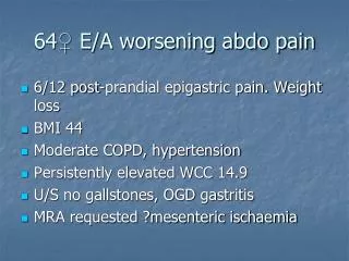
64♀ E/A worsening abdo pain • 6/12 post-prandial epigastric pain. Weight loss • BMI 44 • Moderate COPD, hypertension • Persistently elevated WCC 14.9 • U/S no gallstones, OGD gastritis • MRA requested ?mesenteric ischaemia
MRA • Coeliac occlusion • SMA 95% stenosis • IMA occlusion • 5.2cm AAA • LCIA stenosis 75% • RCIA stenosis 85%
Atrophic right kidney Stenosis origin left renal artery CT
Options? • Percutaneous stent SMA • Access? • Back up plan/ bail out? • Need for subsequent AAA repair • Open repair and mesenteric, renal revascularisation • Iliac angioplasty, hybrid AAA repair with revascularisation
Emergency! • Hypotension • Worsening abdominal pain • Rising WCC 31 • Renal impairment • No peritoneal signs • CT- no rupture AAA, no perforation/free fluid • ITU sepsis ?source • resuscitation. Would not survive major open procedure • Transbrachial angioplasty & stent
Post SMA stent • Abdominal pain improved • Improved gases and WCC halved • No peritonism • Acutely ischaemic arm! • Brachial thrombectomy and patch
Day 3 • Diarrhoea • Rise in WCC, abdominal tenderness • Gases normalised BE -0.6, lactate normal • CT performed • still deemed high anaesthetic risk for laparoscopy/laparotomy
Day 8 • Vomiting overnight, increasing pain • Rise in inflammatory markers • BE -6.3 • ?occluded stent • Duplex- stent patent, dilated SB loops • CT
Laparotomy • Multiple small areas infarction terminal ileum. Remainder very well perfused • Infarcted segment left lobe liver • Ischaemic GB • Ileal resection, cholecystectomy
Discussion • Awareness of possible diagnosis essential • Definitive diagnosis often only made when advanced complications and clear clinical signs • Revascularisation often possible with angioplasty/stent/surgery • Need for vigilance post reperfusion to detect non viable bowel • CT/ relook laparotomy • Stormy post operative course normal • Mucosal sloughing/ loss of gut barrier/ need for parenteral nutrition