Comprehensive Practical Radiology Session for Liver, Spleen, Pancreas, and Biliary System
Explore X-ray evaluation of liver, spleen, kidneys, and psoas muscles, along with ultrasound assessment of liver parenchyma, diaphragm, and biliary structures. Understand CT imaging of abdomen highlighting key structures like gallbladder, pancreas, and blood vessels. MRI insights into stomach, IVC, spleen, liver segments, and gallbladder anatomy.

Comprehensive Practical Radiology Session for Liver, Spleen, Pancreas, and Biliary System
E N D
Presentation Transcript
Practical Radiology Session Liver, spleen, pancreas and biliary system.
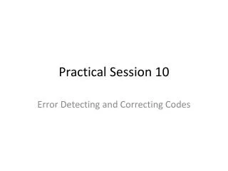
X-ray liver spleen. Right kidney. Left kidney. Right psoas muscle. Left psoas muscle.
US liver LP-liver parenchyma D-diaphragm K-right kidney
US biliary C G- gall bladder P- portal vein C-common bile duct
CT Abdomen 2 8 7 3 6 1 5 4 1-spleen 2-left liver lobe 3-right liver lobe 4-abdominal aorta 5-IVC. 6-pancreatic tail 7-pancreatic body 8-pancreatic head.
CT Abdomen 4 6 1 2 7 3 5 1-gall bladder 2-duodanum 3-pancreatic head 4-SMV 5-SMA 6-CBD 7-PV.
CT Liver PV
MRI Abdomen 17-stomach 21-IVC 30-spleen 1,2,4A, 7,8 liver segments
MRCP 1 5 6 4 2 3
MRCP Cystic duct Gall bladder