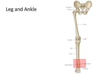
Knee and Ankle
Knee and Ankle. Mazyad Alotaibi. Testing The Muscles of Knee . Knee Extension. ANATOMY: Prim mover /agonist: Quadriceps muscle group which consists of 4 muscles rectus femoris vastus lateralis vastus intermedius vastus medialis Synergist / Accessory Muscles: Tensor Fascia Latae

Knee and Ankle
E N D
Presentation Transcript
Knee and Ankle MazyadAlotaibi
Knee Extension ANATOMY: Prim mover /agonist: Quadriceps muscle group which consists of 4 muscles • rectus femoris • vastuslateralis • vastusintermedius • vastusmedialis Synergist / Accessory Muscles: Tensor Fascia Latae Range of motion: 135 to 0 Substitution: when pt in side-lying (grade 2),pt may use hip internal rotators.
Knee Extension Effect of weakness and contracture: effect of weakness: - difficulty in getting up & down from sitting position as well as on going up & down stairs. - knee hyperextension. effect of contracture : - restriction of knee flexion • shortness of the Rectus Femoris, results in a restriction of the knee flexion when the hip is extended or a restriction of the hip extension when the knee is flexed. Procedures: a- Position of Patient: b- Position of Therapist : inner hand, Outer hand, Direction of Resistance c- Test d- Instruction to patient
Knee Flexion ANATOMY: • Prim mover /agonist: Hamstring muscles Biceps femoris, Semitendinosus, and Semimembranosus - Synergist / Accessory Muscles: Gracilis, TFL, Sartorius, Poplieus, Gastrocnemius, &Plantaris. Range of motion: O to 135 Substitution: - hip flexors. - Sartorius - Gracilis
Knee Flexion Effect of weakness and contracture: effect of weakness: - weakness of both med & lat hamstring causes knee hyperextension. -weakness of lat. Hamstring causes loss of lateral knee stability. -weakness of med. Hamstring decrease medial knee stability. effect of contracture: - knee flexion deformity accompanied by posterior tilting of the pelvis and flattening of the lumbar curve. - restriction of knee extension when the hip is flexed or restriction of the hip flexion when the knee is extended. Procedures: a- Position of Patient: b- Position of Therapist : inner hand, Outer hand, Direction of Resistance c- Test d- Instruction to patient
Knee flexion should be measured with the subject supine. This position allows assessment of the joint range of motion without interference from tightness in the rectus femoris muscle. If the examiner wishes to assess length of the rectus femoris, have the patient lie prone
Ankle Planter Flexion ANATOMY: Prim mover /agonist: Gastrocnemius and Soleus Synergist / Accessory Muscles: Tibialis posterior, plantaris, peroneuslongus & brevis, Flexor digitorum & hallucislongus. Range of motion: 0 to 45 Substitution: • Flexor hallucislongus and flexor digitorumlongus • Peroneuslongus and brevis. • Tibialis posterior.
Ankle Planter Flexion Effect of weakness and contracture: effect of weakness : - result in an hyperextension of the knee as well as in a non-weight bearing position as in standing. - during walking the inability to rise on toes. effect of contracture: result in an equnus position of the foot and flexion of the knee. - also a restriction of the ankle dorsiflexion when the knee is extended and a restriction of the knee extension when the ankle is dorsiflexed. Procedures: WB test and Non WB test a- Position of Patient: b- Position of Therapist : inner hand, Outer hand, Direction of Resistance c- Test d- Instruction to patient
Foot Dorsiflexion and Inversion. ANATOMY: Prime mover/agonist: Tibialis Anterior Synergist/ Accessory muscles: peroneustertius, extensor digitorum and hallucislongus. Range of motion: 0 – 20 Substitution: By the extensor digitorum and extensor hallucislongus muscles results in toes extension
Foot Dorsiflexion and Inversion. Effect of weakness/contracture/shortening: effect of weakness: decrease the ability to dorsiflex the ankle joint (droop foot). effect of contracture: in ability to plantarflex the ankle.
Foot Dorsiflexion and Inversion. Procedures: a- Position of Patient: b- Position of Therapist : inner hand, Outer hand, Direction of Resistance c- Test d- Instruction to patient • Patient Position: • · Sitting, with the patient’s heel resting on the thigh of the therapist. Patient's foot should be dorsiflexed and inverted. • Therapist and Patient Instructions: • · Therapist is sitting on stool beside the limb being tested. The heel of the patient can be resting on the therapist’s thigh. The resistance hand should be placed around the dorsum and medial aspect of the foot. Resistance is given down and out toward eversion. The stabilizing hand is around the posterior leg just above the
Foot Inversion From Planter Flexion ANATOMY: Prime mover/agonist: Tibialis posterior peroneustertius (with Dorsiflexion), extensor digitorum and hallucislongus. Range of motion: 0 – 35 Substitution: the flexors digitorum and hallucislongus muscles results in toes flexion
Foot Inversion From Planter Flexion Effect of weakness/contracture/shortening: effect of weakness:may dropping in medial arch of the foot. ( flat foot). effect of contracture: in ability to plantarflex & evert the ankle. Procedures: a- Position of Patient: b- Position of Therapist : inner hand, Outer hand, Direction of Resistance c- Test d- Instruction to patient
Foot Eversion From Plantar Flexion ANATOMY: • Prime mover/agonist: Peroneuslongus and Peroneusbrevis • Synergist/ Accessory muscles: Extensor digitorumlongus and PeroneusTertius. - Range of motion: 0- 35 - Substitution: No substitution.
Synergist/ Accessory muscles: Extensor digitorumlongus and PeroneusTertius.
Foot EversionfromPlantarFlexion Effect of weakness/contracture/shortening: effect of weakness:may results in: - Decrease the strength of eversion of the foot & planter flexion of the ankle jt. - Decrease lateral stability of the foot. effect of contracture: results in an everted or valgus position of the foot. Procedures: a- Position of Patient: b- Position of Therapist : inner hand, Outer hand, Direction of Resistance c- Test d- Instruction to patient
Test for Lower limb discrepancy Definition: Lower limb discrepancy is a condition of unequal long or round measurements of the lower limbs. Long measurements or Leg length discrepancy (LLD) may comes from difference in length of the femur, or tibia, or both. In some conditions, the whole side is affected, including the upper limbs. Round measurement or Leg circumference discrepancy may comes from difference in lower limbs size due to muscle atrophy or swelling. Leg length discrepancy : Many people walk around with LLD’s of up to 2 cm. and not even know it. However, discrepancies above 2 cm. becomes more noticeable, and a slight limp is present. But even up to 3 cm. a small lift compensates very well. Beyond 3 cm. however, the limp is quite pronounced, and medical care is often sought at that point. Walking with a short leg gait is not only unsightly, but increases energy expenditure during ambulation. It could also put more stress on the long leg, and causes functional scoliosis.
Test for Leg Length Discrepancy A- True leg length Discrepancy Measure from A.S.I.S to inferior border of medial malleolusFix point to fix point • Patient lies supine on a table with trunk, pelvis, and legs in straight alignment and legs close together. The distance from ASIS to the medial malleolus is measured on right and left. • To determine the reason of shorted leg whether in the tibia or in the femur : Ask the patient lies in crock lying position by support his feet on the surface of table with knees and hips flexed. Make good observation on the knees level. (tibia and femur bone). If one knee project further anteriorly than the other, the femur of that extremity is longer. If one knee appears higher than the other, the tibia of that extremity is longer.
Gross Leg Length Discrepancy Magee 4th Edition – pg. 628
B- Apparent leg length discrepancy In apparent shortening, the limb is not altered in length, but appears shortened. This may be as a result of an adduction contracture of the hip joint, which has to be compensated for by tilting of the pelvis, or SIJ pathology causing pelvic rotation. Non-Fix point to fix point -Measure from tip of xiphoid process to inferior border of medial malleolus. - Measure from the umbilicus to the medial malleolus
Leg Length Measurements Eyeball method Measurement method
Measurement of circumference of the knee Swelling around the knee joint • - at middle of patella • -below the patella 5cm • -above the patella 5cm Muscle atrophy: • Above the patella 10cm • Above the patella 15cm • Above the patella 20cm • Above the patella 25cm