Download
1 / 23
230 likes | 361 Vues
Construction of a Microsatellite-Enriched Genomic Library of Physalis philadelphica. Maria Chacon. March 19 2003. Purpose.

E N D
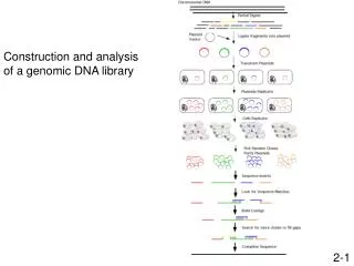
Construction of a Microsatellite-Enriched Genomic Library of Physalis philadelphica Maria Chacon March 19 2003
Purpose Present progress on building a Physalis philadelphica genomic library with a high proportion of inserts containing microsatellite repeats. The protocol used was modified from the one developed at the Natural History Museum of the Smithsonian Institution
Outline • Microsatellite definition and mutation process • Application of microsatellite markers • Advantages of microsatellites • Drawbacks of microsatellites • Protocol • Results • Conclusions and future work
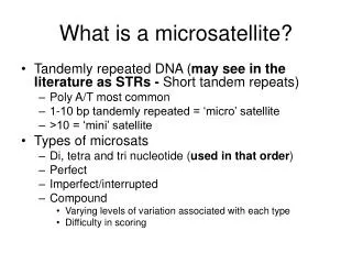
1. Microsatellite definition and mutation process Also known as simple sequence repeats (SSR) or short tandem repeat (STR). These terms are used to describe tandem repeats of short sequence motifs from mono to penta-nucleotides. Classification: Mono (A)11: AAAAAAAAAAA Di (AT)8: ATATATATATATATAT Tri (ATC)7: ATCATCATCATCATCATCATC Tetra (CTAG)6 CTAGCTAGCTAGCTAGCTAGCTAG Imperfect microsatellite GTGTGTGTATGTGTGT Interrupted microsatellite GTGTGTGTCCCGTGTGTGT Compound microsatellite GTGTGTGTCTCTCTCTCTCT
Genomic distribution of microsatellites They are abundant in the eukaryotic genome and are distributed throughout the genome The genomic frequency of microsatellites is inversely related to their repeat number, the higher number of repeats the less frequent Microsatellites not based on a unit of three are rare within coding sequences as these can give rise to frameshift if they mutate
A microstellite mutation model Microsatellite are exposed to a mutational process called DNA (replication) slippage: this causes length instability of tandem repeats and generates polymorphisms (after Schlotterer and Harr, 2001)
2. Applications of microsatellite markers Several hundreds of microsatellites are present in eukaryotic genomes and each locus is subjected to DNA slippage they are therefore a huge reservoir for polymorphic genetic markers Population genetic studies of natural populations: Hybridization, population history and phylogeography, divergence among populations, inbreeding, conservation genetics Behavioral ecology: male mating success determined by paternity testing, social organization of populations (identification of relatedness) and multiple paternity Genetic mapping: Microsatellites are distributed more or less evenly throughout the genome which makes them appropriate markers for mapping
3. Advantages of microsatellites • They probably exist in most of the species • They are codominant markers • They occur throughout most species’ genomes • They can be isolated through the construction of a genomic library enriched for microsatellites or by the use of primers originally design for related species • High heterozygosity level and high mutation rate • Once isolated, microsatellites are amplified by PCR. Multiplex amplification of up to five loci is possible in a single PCR reaction which makes the scoring of multiple genotypes faster and cheaper
4. Drawbacks of microsatellites • Some organisms are very difficult to obtain microsatellite from: Some plants, invertebrates such as Lepidopterans and birds • Problems associated with PCR: A. non-amplification of certain alleles due to substitutions, insertions or deletions within the priming sites generating “null alleles” B. Taq polymerase may generate slippage products or add an extra dNTP which cause single base shifts making typing difficult • Problems associated with size or length homology: alleles may converge on the same size via different types of events in or surrounding the repeat array. This has limited their use in resolving evolutionary relationships
Size or length homoplasy Addition or deletion of another type of repeat unit within the array Nonrepeated sequences or a partial repeat within the array Changes in the sequence flanking the array Five SSR markers in Poplar. Tree Genetic Engineering Research Cooperative Six bovine SSR markers. Kovar et al. LI-COR environmenral products
5. Protocol • Digestion of genomic DNA • Ligation of adapters • Enrichment steps with biotin-labeled SSR probes • Removal of adapters • Cloning of enriched fragments • PCR amplification of inserts • Sequencing of inserts and design of primers
5’-GTTCGATTGCGGATCCTCCTATTAGGATCCCGATCTGA-3’ 3’-CAAGCTAACGCCTAGGAGGATAATCCTAGGGCTAGACT-5’ overhang overhang GTTCGATTGCG CAAGCTAACGCCTAG GATCCCGATCTGA GGCTAGACT GATCCTCCTATTAG GAGGATAATCCTAG overhang overhang Digestion of genomic DNA Genomic DNA is fragmented by digestion with restriction Endonucleases. These are enzymes that cut the DNA at specific recognition sequences DNA was extracted from young leaves of Physalis philadelphica and restricted with BamHI
5’-GCGGTACCCGGGAAGCTTGG 3’- CGCCATGGGCCCTTCGAACCCTAG GATCCCAAGCTTCCCGGGTACCGC-3’ GGTTCGAAGGGCCCATGGCG-5 II. Ligation of adapters Adapters are short DNA fragments of known sequence that may or not contain at the 3’ end an overhang for a specific restriction enzyme. Adapters are linked to both ends of each fragment generated by restriction digestion Adapters help manipulate the digested fragments of unknown sequence GATCCTCCTATTAG GAGGATAATCCTAG BamHI recognition sequences are restored at both ends of restriction fragments
magnet Biotin-labeled SSR probes Straptividine beads (AAC)10, (AAG)10, (AAT)10, (ACT)10, (AGT)10 (ATG)10, (ATC)10, (TTC)10, (TTA)10, (TTG)10 III. Enrichment steps with biotin-labeled SSR probes The purpose of this step is to select the fragments containing microsatellite sequences Adapter-ligated fragments are hybridized with biotin-labeled SSR probes Fragments that do not hybridize with probes are washed away by attaching biotin to straptividine beads and a magnet
IV. Removal of adapters Adapters are removed by digesting with BamHI enzyme. The BamHI overhangs are restored at both ends of the fragments. These overhangs are going to complement overhangs of the vector for cloning 5’-GCGGTACCCGGGAAGCTTGG 3’- CGCCATGGGCCCTTCGAACCCTAG GATCCCAAGCTTCCCGGGTACCGC-3’ GGTTCGAAGGGCCCATGGCG-5 GATCCTCCAACAACAACAACAACTATTAG GAGGTTGTTGTTGTTGTTGATAATCCTAG
BamHI restriction Transformation V. Cloning of insert DNA Linear vector lacZ’ Ampicillin MCS pBluescript vector Ligation of insert Circular vector
Growing in selective medium Transformation of XL1-blue strain of E. Coli pBluescript vector carries a partial copy of the lacZ gene and F’ episome also carries a defective lacZ gene which complement each other to produce an active B-galactosidase gene The active gene gives a blue color. The inactive gene gives a white color Ampr lacZ Tetr lacZ E. coli+plasmid without insert+f’ episome lacZgene Ampr Tetr lacZ E. coli+plasmid with insert+f’episome Ampicillin + Tetracycline + IPTG + X-GAL Ampr E. coli+plasmid without insert, no f’ episome
White-blue color selection Ampr lacZ expression repression inhibited by IPTG X-Gal degrades Functional lacZ gene lac repressor lacZgene X-Gal does not degrade Non-expression Ampr Non-functional lacZ gene
VI. PCR amplification of inserts T7 primer T3 primer
VII. Sequencing of inserts Partial sequence of an insert enriched with (TTG)10
6. Results Enrichment was succesful for all microsatellite probes except for (ATG)10 and (TTC)10 Several clones with insert were obtained for the successful enrichment reactions: (TTG)10: 250 clones (AGT)10: 126 clones (TTA)10: 90 clones (AAC)10: 50 clones (ACT)10: 50 clones (AAT)10: 130 clones (AAG)10+(ATC)10: 72 clones Total: 768 clones
6. Results Insert size ranged from 300 bp up to 1500 bp. The most common sizes ranged from 300-700 bp 33 clones: 10 enriched for (TTG)10, 14 for (AGT)10, 5 for (TTA)10 and 3 for (AAC)10 were sent for sequencing. One third did not contain SSR including all those that were enriched for TTA
Conclusions and future work Physalis philadelphica contains AT-rich microsatellites as other plant species and this method have proved useful for isolating them Microsatellite sequences can be isolated by doing one or two steps of enrichment without need for further screening such as hybridization of clones with SSR-probes Ninety six clones are going to be sequenced The aim is to isolate a minimum of 15-20 polymorphic loci